Observational Study
. 2016 Jan;95(2):e2497.
doi: 10.1097/MD.0000000000002497.

Inhibitory Effects of α-Lipoic Acid on Oxidative Stress-Induced Adipogenesis in Orbital Fibroblasts From Patients With Graves Ophthalmopathy

Affiliations
Observational Study

Inhibitory Effects of α-Lipoic Acid on Oxidative Stress-Induced Adipogenesis in Orbital Fibroblasts From Patients With Graves Ophthalmopathy

Sena Hwang et al. Medicine (Baltimore). 2016 Jan.

Abstract

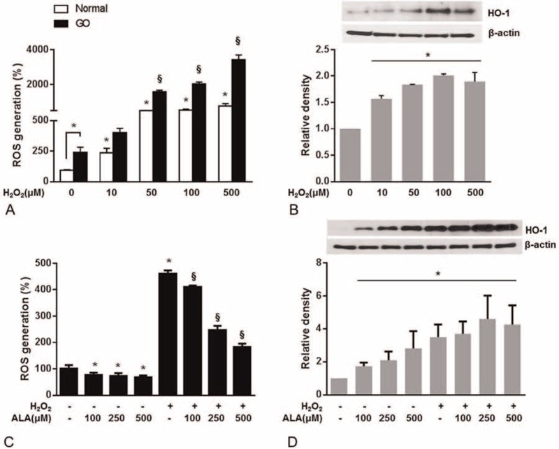
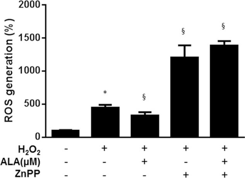
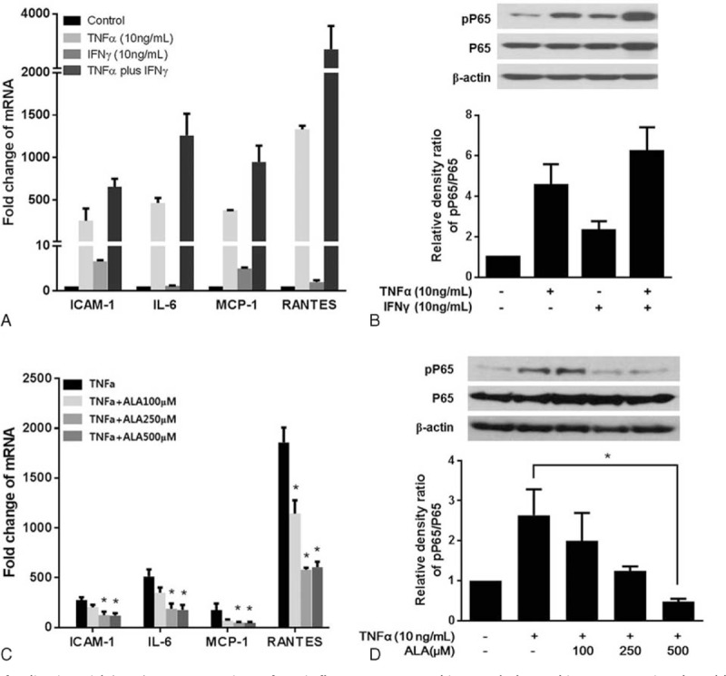
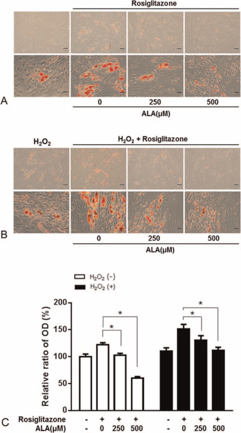
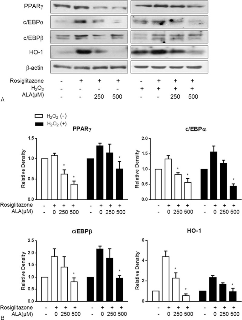
A choice of the optimal treatment for Graves ophthalmopathy (GO) is a challenge due to the complexity of the pathogenesis. Alpha-lipoic acid (ALA) is well known as a multifunctional antioxidant, helping to protect cells against oxidative stress and inflammatory damage.The aim of this study was to investigate the effects of ALA on intracellular production of reactive oxygen species (ROS), inflammation, and adipogenesis using primary cultured orbital fibroblasts from patients with GO.Intracellular ROS levels and mRNA expressions of proinflammatory cytokines and chemokines including intercellular adhesion molecule-1 (ICAM-1), interleukin (IL)-6, monocyte chemoattractant protein (MCP)-1, and regulated upon activation normal T cell expressed and presumably secreted (RANTES) were measured. After adipogenesis, the expressions of peroxisome proliferator-activated receptor (PPAR)γ, CCAAT-enhancer-binding proteins (C/EBP)α and β, and heme oxygenase-1 (HO-1) were investigated.H2O2 dose-dependently stimulated ROS production and HO-1 expression. Addition of ALA strongly attenuated ROS production and further increased HO-1 expression. However, by pretreatment of zinc protoporphyrin (ZnPP), HO-1 inhibitor, ALA inhibition of ROS generation by H2O2 was abolished. Tumor necrosis factor (TNF)α-induced mRNA expressions of ICAM-1, IL-6, MCP-1, and RANTES were inhibited by ALA treatment. In this context, TNFα-induced phosphorylation of P65 was also inhibited. In addition, ALA dose-dependently inhibited H2O2-induced intracellular accumulation of lipid droplets. The expression of adipogenic transcription factors, including PPARγ, C/EBPα, and β, was also inhibited.ALA is a potential therapeutic agent for GO because of the inhibitory effects on ROS production and gene expression of proinflammatory cytokines and chemokines, resulting in prevention of adipose-tissue expansion.


Conflict of interest statement

The authors have no conflicts of interest to disclose.

Figures

FIGURE 1
FIGURE 1
Effects of α-lipoic acid (ALA) on H2O2-induced intracellular production of reactive oxygen species (ROS) and HO-1 expression in fibroblasts from patients with Graves ophthalmopathy (GO). A, Intracellular ROS were quantified by flow cytometry with 5-(and 6)-carboxy-2′,7′-dichlorodihydrofluorescein diacetate (H2DCFDA) in healthy and GO-related cells stimulated with 10 to 500 μM H2O2 for 30 min. B, HO-1 protein expression was determined by western blotting in GO-related orbital fibroblasts stimulated with 0 to 500 μM H2O2 for 24 h. C, The GO-related orbital fibroblasts were treated with ALA (100, 250, or 500 μM) for 24 h and then stimulated with 100 μM H2O2 for 30 min. ROS were quantified by flow cytometry with H2DCFDA. D, HO-1 expression in the GO-related cells was analyzed by western blotting. The GO-related cells were treated with ALA (100, 250, or 500 μM) for 24 h with or without 100 μM H2O2. The results are expressed as a percentage of no-treatment control values, mean ± SEM (n = 3). P < 0.05 compared with untreated cells; §P < 0.05 compared with cells stimulated with H2O2.
FIGURE 2
FIGURE 2
A heme oxygenase 1 (HO-1) inhibitor, zinc protoporphyrin (ZnPP), abrogated the inhibitory effect of α-lipoic acid (ALA) on production of reactive oxygen species (ROS). Orbital fibroblasts from patients with Graves ophthalmopathy (GO) were pretreated with ZnPP (10 μM) for 1 h followed by treatment with ALA (500 μM) for 24 h, and were then stimulated with 100 μM H2O2 for 30 min. ROS were quantified by flow cytometry with 5-(and 6)-carboxy-2′,7′-dichlorodihydrofluorescein diacetate (H2DCFDA). The data are expressed as percentages of the no-treatment control values, mean ± SEM (n = 3). P < 0.05 compared with the untreated cells; §P < 0.05 compared with the cells stimulated with H2O2.
FIGURE 3
FIGURE 3
Effects of α-lipoic acid (ALA) on expression of proinflammatory cytokine and chemokine genes stimulated by TNF-α. A, The cells were incubated with 10 ng/mL TNF-α and 10 ng/mL IFNγ, alone or in combination for 24 h. The mRNA levels were determined by RT-PCR, and the results were normalized to the housekeeping gene, GAPDH, and expressed in arbitrary units relative to the levels of no-treatment control, set to 1.0. B, Phosphorylation of P65 (pP65) in the whole-cell extract was determined by western blotting. Quantification of pP65 and P65 by densitometry, normalized to the level of β-actin in the same sample, is shown. C, The cells were incubated with ALA (0, 100, 250, or 500 μM) and 10 ng/mL TNF-α for 24 h. The mRNA levels were determined by RT-PCR. D, The protein expression of pP65 was determined by western blotting. The data in the columns are mean relative density ratios ± SEM (n = 3). P < 0.05 compared with the cells stimulated with 10 ng/mL TNF-α.
FIGURE 4
FIGURE 4
The effect of α-lipoic acid (ALA) on adipogenesis of orbital fibroblasts from patients with Graves ophthalmopathy (GO). Treatment with ALA (250 or 500 μM) for the first 3 days after initiation of the 10-day adipogenesis procedure in the adipogenic medium with or without 10 μM rosiglitazone (A) or its combination with H2O2 (B). The cells were stained with Oil Red O and examined macroscopically and microscopically (×40 and ×400 magnifications). C, Cell-bound Oil Red O was solubilized, and OD at 490 nm was measured to quantify the adipogenesis. The data in the column are the mean relative density ratios ± SEM (n = 3). P < 0.05 compared with the cells that differentiated under the influence of rosiglitazone or its combination with H2O2 (10 μM).
FIGURE 5
FIGURE 5
Effects of α-lipoic acid (ALA) on the expression of adipogenic transcriptional regulators during adipogenesis of orbital fibroblasts from patients with Graves ophthalmopathy (GO). Western blot analysis of protein expression of PPARγ, c/EBPα, c/EBPβ, and HO-1 (A). Quantification by densitometry, after normalization to the β-actin level in the same sample, is shown for PPARγ (B), c/EBPα (C), c/EBPβ (D), and HO-1 (E). The data in the column are mean relative density ratios ± SEM (n = 3). P < 0.05 compared with the cells that differentiated under the influence of rosiglitazone or its combination with H2O2 (10 μM).

Similar articles

Cited by

References

    1. Bahn RS. Graves’ ophthalmopathy. N Engl J Med 2010; 362:726–738. - PMC - PubMed
    1. Smith T. Pathogenesis of Graves’ orbitopathy: a 2010 update. J Endocrinol Invest 2010; 33:414–421. - PMC - PubMed
    1. Korducki J, Loftus S, Bahn R. Stimulation of glycosaminoglycan production in cultured human retroocular fibroblasts. Invest Ophthalmol Vis Sci 1992; 33:2037–2042. - PubMed
    1. Bartalena L, Tanda ML, Piantanida E, et al. Oxidative stress and Graves’ ophthalmopathy: in vitro studies and therapeutic implications. Biofactors 2003; 19:155–163. - PubMed
    1. Burch HB, Lahiri S, Bahn RS, et al. Superoxide radical production stimulates retroocular fibroblast proliferation in Graves’ ophthalmopathy. Exp Eye Res 1997; 65:311–316. - PubMed

Publication types

MeSH terms