. 2016 May;24(5):965-77.
doi: 10.1038/mt.2016.12. Epub 2016 Jan 14.

Human Mesenchymal Stem Cells Genetically Engineered to Overexpress Brain-derived Neurotrophic Factor Improve Outcomes in Huntington's Disease Mouse Models

Affiliations

Human Mesenchymal Stem Cells Genetically Engineered to Overexpress Brain-derived Neurotrophic Factor Improve Outcomes in Huntington's Disease Mouse Models

Kari Pollock et al. Mol Ther. 2016 May.

Abstract

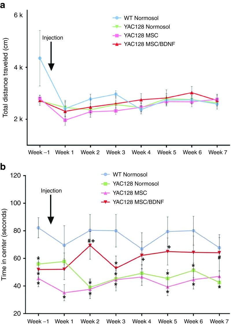
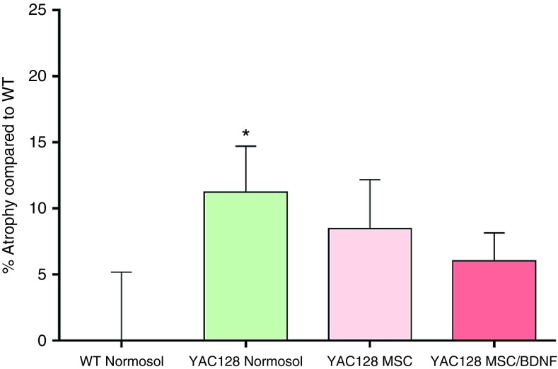
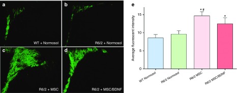
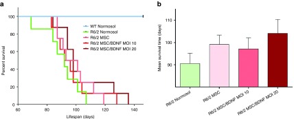
Huntington's disease (HD) is a fatal degenerative autosomal dominant neuropsychiatric disease that causes neuronal death and is characterized by progressive striatal and then widespread brain atrophy. Brain-derived neurotrophic factor (BDNF) is a lead candidate for the treatment of HD, as it has been shown to prevent cell death and to stimulate the growth and migration of new neurons in the brain in transgenic mouse models. BDNF levels are reduced in HD postmortem human brain. Previous studies have shown efficacy of mesenchymal stem/stromal cells (MSC)/BDNF using murine MSCs, and the present study used human MSCs to advance the therapeutic potential of the MSC/BDNF platform for clinical application. Double-blinded studies were performed to examine the effects of intrastriatally transplanted human MSC/BDNF on disease progression in two strains of immune-suppressed HD transgenic mice: YAC128 and R6/2. MSC/BDNF treatment decreased striatal atrophy in YAC128 mice. MSC/BDNF treatment also significantly reduced anxiety as measured in the open-field assay. Both MSC and MSC/BDNF treatments induced a significant increase in neurogenesis-like activity in R6/2 mice. MSC/BDNF treatment also increased the mean lifespan of the R6/2 mice. Our genetically modified MSC/BDNF cells set a precedent for stem cell-based neurotherapeutics and could potentially be modified for other neurodegenerative disorders such as amyotrophic lateral sclerosis, Alzheimer's disease, and some forms of Parkinson's disease. These cells provide a platform delivery system for future studies involving corrective gene-editing strategies.


Figures

Figure 1
Figure 1
Vector map and stability. (a) Vector map diagram for pCCLc-MNDU3-BDNF-WPRE. A third-generation lentiviral vector, based on the pCCLc-x backbone was used to generate a BDNF gene construct. The transgene is driven by the MNDU3 promoter, a constitutive RNA polymerase II promoter. BDNF is the gene encoding brain-derived neurotrophic factor, cloned in our lab from adult human bone marrow-derived MSCs. WPRE is the woodchuck hepatitis post-transcriptional regulatory element, which acts in cis to enhance gene expression. (b) Stability of the clinical vector in transduced cells. To determine whether there were any deletions or rearrangements in vector-transduced cells, genomic PCR was performed. MSCs were transduced with the BDNF vector at MOI 10 or MOI 20. Total genomic DNA was extracted and analyzed by PCR with primers specific for the respective vector segments. (I) ψ (forward) and U5-3' (reverse), (II) ψ (forward) and MNDU3 (reverse), (III) MNDU3 (forward) and BDNF (reverse), and (IV) BDNF (forward) and U5-3' (reverse). Resulting DNA was run on a gel in the following lanes: 1 kb DNA ladder (DLa), nontransduced MSCs (negative control, lane 1), BDNF MOI 10 MSCs (lane 2), BDNF MOI 20 MSCs (lane 3), BDNF vector plasmid (positive control, lane 4), no template control (lane 5). Bands were of the expected size, as shown in the schematic of the PCR products below the panels. Ψ, psi packaging sequence; BDNF, brain-derived neurotrophic factor; WPRE, woodchuck hepatitis virus post-transcriptional regulatory element.
Figure 2
Figure 2
Cell characterization. (a) Cells transduced by the BDNF vector (right), display the size and phenotypic appearance of healthy nontransduced MSCs from the same donor (left). (b) In vitro characterization showed that human MSCs transduced with the pivotal BDNF lentiviral vector at multiplicities of infection (MOI) 2, 5, 10, and 20 displayed a positive correlation between BDNF production and MOI. Supernatant was collected for determination of secreted BDNF by ELISA after conditioning serum free media for 24 hours. (c) Vector copy number at increasing MOI. Human MSCs were transduced with pCCLc-MNDU3-BDNF-WPRE, cryopreserved, then subsequently thawed and expanded for 3 days. DNA was isolated and used for qPCR analysis. Quantification was based on standard curves of plasmid DNA. Vector copy number/cell was determined as WPRE/2GAPDH.
Figure 3
Figure 3
Cell differentiation and proliferation. (a) Differentiation potential of MSC is not affected after genetic modification by the BDNF vector. Osteogenesis was assessed by Alizarin Red Staining of precipitated calcium after 16 days in culture in osteogenic media. Representative images (10×) are shown. Adipogenesis was assessed after 16 days in adipogenic media by formation of adipocytes using Oil Red O to stain triglycerides. Representative images (10×) are shown. (b) Proliferation rates were not significantly altered in MSCs post-transduction. Passage 6 MSCs were plated at approximately 1,000 cells/cm2 in 24-well plates and cultured as described. After 1, 3, 5, 7, and 9 days in culture, plates were analyzed using CellTiter96 nonradioactive cell proliferation assay (Promega).
Figure 4
Figure 4
In vivo imaging demonstrates that immune suppression (of FVB/NJ mice) was able to increase cell retention to levels similar to that observed in immune-deficient NSG mice for at least 28 days. Human MSCs were transduced by a lentiviral vector carrying the luciferase gene (pCCLc-MNDU3-Luc-PGK-EGFP-WPRE), which allows cells to be visualized (bioluminescence) in the brains of living mice over time.
Figure 5
Figure 5
Open-field analysis to measure anxiety. (a,b) Each animal was placed into an open arena once a week and monitored for 10 minutes. All data was collected using Fusion system software. Total distance traveled is a measure of spontaneous exploration and is an indicator motor ability. The time in the center zone of the box was measured to test anxiety. * = Significant to WT; # = Significant to Tg + Normosol; † = Significant to Tg + MSC; n = 16–17/group.
Figure 6
Figure 6
Striatal atrophy. Following completion of behavioral analysis in the YAC128 efficacy study, animals were euthanized and formalin perfused. Brains were cryosectioned at 30 μm and labeled with cytochrome oxidase. Brains were imaged using a Keyence BZ-9000 microscope and striatal volume was calculated using the Cavalieri principle for volume estimation. Striatal atrophy was calculated by first normalizing striatal volume to the WT group, then subtracting the value from 100. * = Significant to WT.
Figure 7
Figure 7
Neurogenesis. Three weeks following transplantation of MSC, MSC/BDNF MOI 10, or vehicle control (Normosol-R), the 10-week-old R6/2 mice were euthanized and formalin perfused. Brains were cryosectioned at 30 μm and labeled using doublecortin and AlexaFluor488. The subventricluar zone was imaged using a Zeiss Axioskop 2 microscope (10× objective) and average fluorescent intensity was calculated using ImageJ. * = Significant to WT, # = Significant to R6/2 + Normosol. n = 6–7/group.
Figure 8
Figure 8
Implantation of MSC/BDNF increased the lifespan of R6/2 mice. Kaplan-Meier analysis: 10% increase for WT MSC, 7.7% increase for MSC/BDNF MOI 10, 15.5% increase for MSC/BDNF MOI 20. n = 7–9 mice per group.

Similar articles

Cited by

References

    1. A novel gene containing a trinucleotide repeat that is expanded and unstable on Huntington's disease chromosomes. The Huntington's Disease Collaborative Research Group. Cell 72: 971–983 (1993). - PubMed
    1. Huntington Study, G (2006). Tetrabenazine as antichorea therapy in Huntington disease: a randomized controlled trial. Neurology 66: 366–372. - PubMed
    1. Killoran, A and Biglan, KM (2014). Current therapeutic options for Huntington's disease: good clinical practice versus evidence-based approaches? Mov Disord 29: 1404–1413. - PubMed
    1. Scheuing, L, Chiu, CT, Liao, HM, Linares, GR and Chuang, DM (2014). Preclinical and clinical investigations of mood stabilizers for Huntington's disease: what have we learned? Int J Biol Sci 10: 1024–1038. - PMC - PubMed
    1. Canals, JM, Pineda, JR, Torres-Peraza, JF, Bosch, M, Martín-Ibañez, R, Muñoz, MT et al. (2004). Brain-derived neurotrophic factor regulates the onset and severity of motor dysfunction associated with enkephalinergic neuronal degeneration in Huntington's disease. J Neurosci 24: 7727–7739. - PMC - PubMed

Publication types