. 2016 Jun;31(6):1287-99.
doi: 10.1002/jbmr.2787. Epub 2016 Mar 15.

Class I and IIa HDACs Mediate HIF-1α Stability Through PHD2-Dependent Mechanism, While HDAC6, a Class IIb Member, Promotes HIF-1α Transcriptional Activity in Nucleus Pulposus Cells of the Intervertebral Disc

Affiliations

Class I and IIa HDACs Mediate HIF-1α Stability Through PHD2-Dependent Mechanism, While HDAC6, a Class IIb Member, Promotes HIF-1α Transcriptional Activity in Nucleus Pulposus Cells of the Intervertebral Disc

Zachary R Schoepflin et al. J Bone Miner Res. 2016 Jun.

Abstract

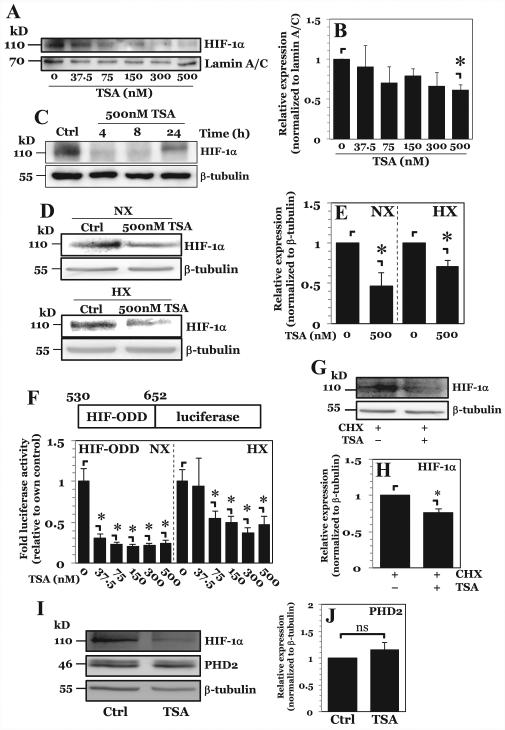
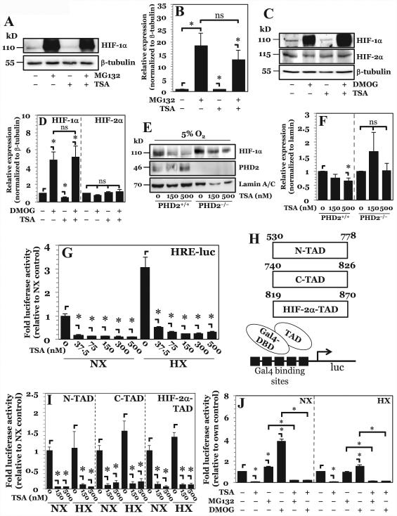
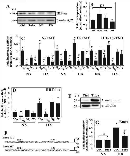
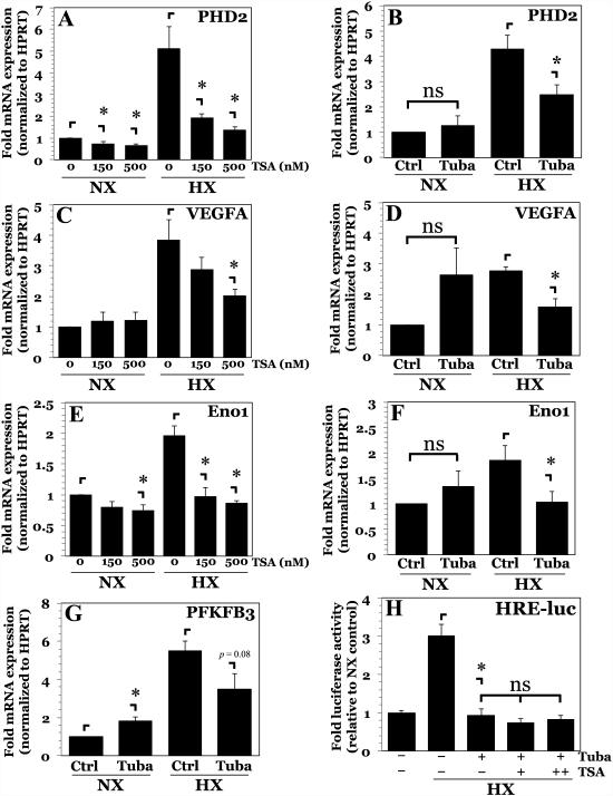
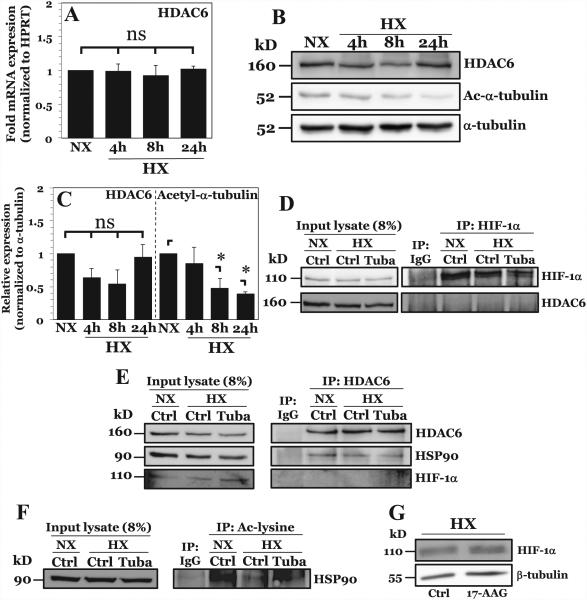
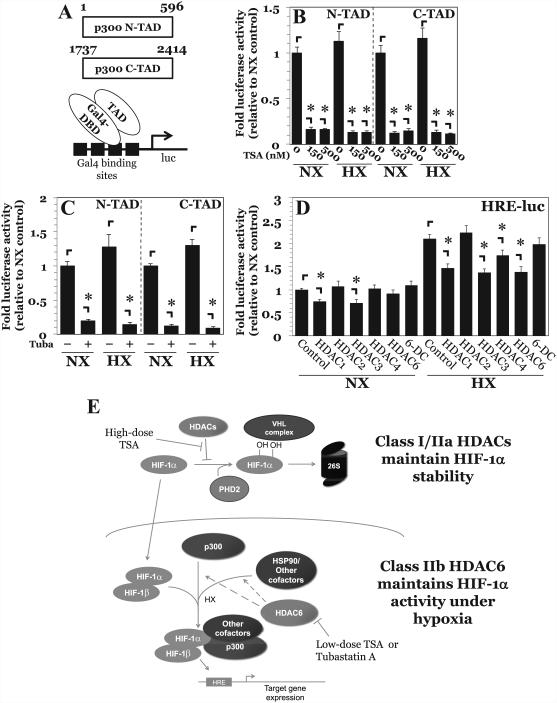
The objective of this study was to determine the role of histone deacetylases (HDACs) in regulating HIF-1α protein stability and activity in nucleus pulposus (NP) cells. Treatment of NP cells with pan-HDAC inhibitor TSA resulted in decreased HIF-1α levels under both normoxia and hypoxia in a dose-dependent fashion. TSA-mediated HIF-1α degradation was rescued by concomitant inhibition of not only the 26S proteasome but also PHD2 function. Moreover, TSA treatment of PHD2(-/-) cells had little effect on HIF-1α levels, supporting the notion that inhibition of PHD2 function by HDACs contributed to HIF-1α stabilization. Surprisingly, class-specific HDAC inhibitors did not affect HIF-1α protein stability, indicating that multiple HDACs controlled HIF-1α stability by regulating HIF-1α-PHD2 interaction in NP cells. Interestingly, lower-dose TSA that did not affect HIF-1α stability decreased its activity and target gene expression. Likewise, rescue of TSA-mediated HIF-1α protein degradation by blocking proteasomal or PHD activity did not restore HIF-1 activity, suggesting that HDACs independently regulate HIF-1α stability and activity. Noteworthy, selective inhibition of HDAC6 and not of class I and IIa HDACs decreased HIF-1-mediated transcription under hypoxia to a similar extent as lower-dose TSA, contrasting the reported role of HDAC6 as a transcriptional repressor in other cell types. Moreover, HDAC6 inhibition completely blocked TSA effects on HIF-1 activity. HDAC6 associated with and deacetylated HSP90, an important cofactor for HIF-1 function in NP cells, and HDAC6 inhibition decreased p300 transactivation in NP cells. Taken together, these results suggest that although multiple class I and class IIa HDACs control HIF-1 stability, HDAC6, a class IIb HDAC, is a novel mediator of HIF-1 activity in NP cells possibly through promoting action of critical HIF-1 cofactors. © 2016 American Society for Bone and Mineral Research.

Keywords: HDAC6; HIF-1α; HYPOXIA; INTERVERTEBRAL DISC; NUCLEUS PULPOSUS; TRANSCRIPTION FACTOR.


Figures

Figure 1
Figure 1. Pan-HDAC inhibition in NP cells decreases HIF-1α protein stability in a dose dependent fashion
A, B) Western blot analysis of HIF-1α (A) and corresponding densitometric quantification of at least 3 independent experiments (B) following exposure of NP cells to increasing doses of pan-HDAC inhibitor Trichostatin A (TSA) under hypoxia. Level of nuclear HIF-1α protein decreases only at highest dose of 500 nM of TSA. C) Western blot analysis of HIF-1α after higher-dose TSA treatment for 4-24 hours. D, E) Western blot analysis (D) and corresponding densitometric quantification of at least 3 independent sets (E) of HIF-1α after 8hr higher-dose TSA treatment under both normoxia and hypoxia. TSA treatment decreases HIF-1α expression under both conditions. F) Stability of HIF-ODD-luciferase fusion construct after increasing doses of TSA under both normoxia and hypoxia. G, H) Western blot (G) and corresponding densitometry (H) of HIF-1α in NP cells after 30 minute pretreatment with cycloheximide (CHX, 50 μg/mL) followed by treatment with 500 nM TSA. TSA treatment still results in increased HIF-1α degradation in the presence of CHX. I, J) Western blot (I) and corresponding densitometry (J) of PHD2 in NP cells after TSA treatment showing no change in PHD2 expression upon pan-HDAC inhibition. Quantitative data is represented as mean ± S.E. of three independent experiments performed in triplicate (n = 3); *, p < 0.05.
Figure 2
Figure 2. Higher-dose TSA treatment results in increased HIF-1α turnover through PHD-26S proteasome pathway while lower-dose TSA treatment independently decreases HIF-1α activity
A, B) Western blot analysis of HIF-1α (A) and corresponding densitometric quantification of at least 3 independent experiments (B) after treatment of NP cells with either proteasomal inhibitor MG132 (10 μM) or TSA (500 nM), or both. Decrease in HIF-1α protein seen with higher-dose TSA treatment is rescued by proteasomal inhibition. C, D) Western blot analysis (C) and corresponding densitometry (D) of HIF-1α in NP cells after exposure to PHD inhibitor dimethyloxalylglycine (DMOG, 2 mM) TSA (500 nM), or both. Decrease in HIF-1α protein levels seen with higher-dose TSA treatment is rescued by PHD inhibition. E, F) Western blot (E) and corresponding densitometry of at least 3 independent blots (F) of HIF-1α in PHD2+/+ and PHD2−/− MEFs after exposure to TSA. HIF-1α shows no degradation following higher-dose TSA treatment in PHD2−/− cells. G) HRE-luciferase reporter activity under both normoxia and hypoxia following treatment with increasing doses of TSA. HRE activity decreases with all doses of TSA irrespective of oxygen tension. H) Schematic of Gal-4-TAD constructs and the assay system used for studies described in (I). I) HIF-1α-N-TAD, -C-TAD, and HIF-2α-TAD activity under normoxia and hypoxia after TSA treatment. Both lower dose and higher dose TSA decrease TAD activity under both normoxia and hypoxia. J) HRE-luc activity in NP cells under NX or HX after treatment with MG132, DMOG, and TSA. Data is represented as mean ± S.E. of three independent experiments performed in triplicate (n = 3); *, p < 0.05.
Figure 3
Figure 3. HDAC6 inhibition results in decreased HIF-1α activity independent of effects on protein stability
A, B) Western blot of HIF-1α (A) and corresponding densitometric analysis of multiple independent experiments (B) after NP cell treatment with inhibitors Tubastatin A (15 μM), MC1568 (20 μM), or PD-106 (10 μM). Selective inhibition of HDAC6, class IIa HDACs, or class I HDACs had no effect on HIF-1α protein stability. C, D) HIF-1α-N-TAD, HIF-1α-C-TAD, and HIF-2α-TAD and D) HRE-luciferase reporter activity activity under normoxia and hypoxia after Tubastatin A, MC1568, or PD-106 treatment. A decrease in HIF-1α-N-TAD and HIF-1α-C-TAD activity was seen with inhibition of HDAC6 and Class I HDACs. Endogenous HIF-1α activity, however, was decreased only by HDAC6 inhibition under both normoxia and hypoxia. E) Western blot analysis of α-tubulin, a known target of HDAC6, shows significantly enhanced acetylation (Ac-α-tubulin) after treatment with Tubastatin A. F) Schematic of enolase-1 promoters used in studies. G) Enolase promoter activity, normalized to activity of promoter with mutated HRE sites, after treatment with Tubastatin A under normoxia and hypoxia. Effects of HDAC6 inhibition on promoter activity are only seen under hypoxia. Data is represented as mean ± S.E. of three independent experiments performed in triplicate (n = 3); *, p < 0.05.
Figure 4
Figure 4. Inhibition of HDAC6 in NP cells abrogates hypoxic induction of HIF-1 target genes
A, B) mRNA expression of PHD2 measured by qPCR after TSA (A) or Tubastatin A (B) treatment. C, D) mRNA expression of VEGFA measured by qPCR after TSA (C) or Tubastatin A (D) treatment. E, F) mRNA expression of Eno1 measured by qPCR after TSA (E) or Tubastatin A (F) treatment. G) mRNA expression of PFKFB3 measured by qPCR after Tubastatin A treatment. H) HRE-reporter activity in NP cells pretreated with Tubastatin A following addition of increasing doses of TSA (37.5-150 nM) under hypoxia. Data is represented as mean ± S.E. of three independent experiments performed in triplicate (n = 3); *, p < 0.05.
Figure 5
Figure 5. HDAC6 promotes HIF-1 activity in NP cells under hypoxia through positive effects on HIF-1 transcriptional cofactor HSP90
A) mRNA expression of HDAC6 measured by qPCR after culture in HX for 4-24 hours. B, C) Western blot analysis of HDAC6 and acetylated α-tubulin (Ac-α-tubulin) (B) and corresponding densitometric quantification of experiments (C) following NP cell culture in hypoxia (HX) for 4-24 h. HDAC6 activity, but not expression, increases under hypoxia. D) Immunoprecipitation of HIF-1α in NP cells under normoxia and hypoxia. HDAC6 was unable to be co-precipitated with HIF-1α. E) Immunoprecipitation of HDAC6 in NP cells with or without Tubastatin A treatment. HSP90, but not HIF-1α, was immunoprecipitated with HDAC6 under both normoxia and hypoxia. F) Immunoprecipitation of acetyl-lysine residues in NP cells under normoxia and hypoxia with or without Tubastatin A. Acetylation of HSP90 decreases under hypoxia and increases with HDAC6 inhibition. G) Western blot analysis of HIF-1α after HSP90 inhibition by 8 hr treatment with 17-AAG (500 nM). HSP90 inhibition results in no appreciable change in HIF-1α protein levels. Quantitative data is represented as mean ± S.E. of three independent experiments performed in triplicate (n = 3); *, p < 0.05.
Figure 6
Figure 6. HDAC6 inhibition decreases p300 activity in NP cells
A) Schematic of p300-TAD constructs and the assay system. B, C) p300-N-TAD and p300-C-TAD activity under normoxia and hypoxia after treatment with TSA (150-500 nM) (B) and Tubastatin A (15 μM) (C). Treatment with either TSA or Tubastatin decreased p300 transactivation potential under both normoxia and hypoxia. D) HRE-luciferase activity after transient overexpression of various HDAC isoforms. Overexpression of HDAC1 and HDAC3 irrespective of oxygen tension, and of HDAC4 and HDAC6 under hypoxia decreased HIF-1 activity. E) Proposed model of regulation of HIF-1α stability and activity by HDACs in NP cells. Multiple Class I and Class IIa HDACs promote HIF-1α stability in NP cells by regulating PHD2-HIF-1 interaction. In contrast, HDAC6, independently of regulating HIF-1α stability, promotes hypoxic induction of HIF-1 activity through positive effects on multiple cofactors including p300 and chaperone HSP90.

References

    1. Shapiro IM, Vresilovic EJ, Risbud MV. Is the spinal motion segment a diarthrodial polyaxial joint: What a nice nucleus like you doing in a joint like this? Bone. 2012;50:771–6. - PMC - PubMed
    1. Gruber HE, Ashraf N, Kilburn J, et al. Vertebral endplate architecture and vascularization: application of micro-computerized tomography, a vascular tracer, and immunocytochemistry in analyses of disc degeneration in the aging sand rat. Spine (Phila Pa 1976) 2005;30:2593–600. - PubMed
    1. Risbud MV, Schipani E, Shapiro IM. Hypoxic regulation of nucleus pulposus cell survival: from niche to notch. Am J Pathol. 2010;176:1577–83. - PMC - PubMed
    1. Johnson ZI, Shapiro IM, Risbud MV. Extracellular osmolarity regulates matrix homeostasis in the intervertebral disc and articular cartilage: Evolving role of TonEBP. Matrix Biol. 2014;40:10–6. - PMC - PubMed
    1. Rajpurohit R, Risbud MV, Ducheyne P, Vresilovic EJ, Shapiro IM. Phenotypic characteristics of the nucleus pulposus: Expression of hypoxia inducing factor-1, glucose transporter-1 and MMP-2. Cell Tissue Res. 2002;308:401–7. - PubMed

MeSH terms

Substances