. 2016 Jan 14;11(1):e0146012.
doi: 10.1371/journal.pone.0146012. eCollection 2016.

New Amniotic Membrane Based Biocomposite for Future Application in Reconstructive Urology

Affiliations

New Amniotic Membrane Based Biocomposite for Future Application in Reconstructive Urology

Jan Adamowicz et al. PLoS One. .

Abstract

Objective: Due to the capacity of the amniotic membrane (Am) to support re-epithelisation and inhibit scar formation, Am has a potential to become a considerable asset for reconstructive urology i.e., reconstruction of ureters and urethrae. The application of Am in reconstructive urology is limited due to a poor mechanical characteristic. Am reinforcement with electrospun nanofibers offers a new strategy to improve Am mechanical resistance, without affecting its unique bioactivity profile. This study evaluated biocomposite material composed of Am and nanofibers as a graft for urinary bladder augmentation in a rat model.

Material and methods: Sandwich-structured biocomposite material was constructed from frozen Am and covered on both sides with two-layered membranes prepared from electrospun poly-(L-lactide-co-E-caprolactone) (PLCL). Wistar rats underwent hemicystectomy and bladder augmentation with the biocomposite material.

Results: Immunohistohemical analysis (hematoxylin and eosin [H&E], anti-smoothelin and Masson's trichrome staining [TRI]) revealed effective regeneration of the urothelial and smooth muscle layers. Anti-smoothelin staining confirmed the presence of contractile smooth muscle within a new bladder wall. Sandwich-structured biocomposite graft material was designed to regenerate the urinary bladder wall, fulfilling the requirements for normal bladder tension, contraction, elasticity and compliance. Mechanical evaluation of regenerated bladder wall conducted based on Young's elastic modulus reflected changes in the histological remodeling of the augmented part of the bladder. The structure of the biocomposite material made it possible to deliver an intact Am to the area for regeneration. An unmodified Am surface supported regeneration of the urinary bladder wall and the PLCL membranes did not disturb the regeneration process.

Conclusions: Am reinforcement with electrospun nanofibers offers a new strategy to improve Am mechanical resistance without affecting its unique bioactivity profile.


Conflict of interest statement

Competing Interests: The authors have declared that no competing interests exist.

Figures

Fig 1
Fig 1. Preparation and structure of biocomposite.
(A) The pieces of Am (black arrows) placed onto a sheet of PLCL nanofibers. A drum is used as a target during the nanofiber production process. SEM images are displayed in B-D. (B) A cross-section image of the biocomposite material. The biocomposite material is 389 um thick with an inner cavity containing the Am. (C) Visible drops of glycerin used for Am preservation are observed on surface of PLCL nanofibers (white arrows). (D) Two pieces of delaminated biocomposite material. The borders between consecutive sheets of nanofibers (*) are clearly visible with Am inside.
Fig 2
Fig 2. Cytotoxity testing of biocomposite using MSC.
(A) MSC migrating towards Am on the 7th day of in vitro cultivation. (B) Clusters of MSC distributed on an external surface of a PLCL membrane. (C) Dispersed MSC adherent to PLCL nanofibers.
Fig 3
Fig 3. Amniotic membrane extract cytotoxicity measurement using the MTT assay.
Each result was presented as an average from 5 independent experiment with SD bars. No statistically significant difference in cell viability was observed between AME treated and control cells (p>0.05) after 24 hours.
Fig 4
Fig 4. Amniotic membrane extract cytotoxicity measurement using real-time cell analysis.
Each result was presented as mean from 5 independent experiment with SD bars. No statistically significant differences in cell viability were observed between AME treated and control cells (p>0.05) after 72 hours.
Fig 5
Fig 5. Urinary bladder augmented with biocomposite.
(A) Biocomposite material scaffold prepared for the suture procedure. (B) Urinary bladder after the augmentation procedure. Single fixing sutures are visible (black arrows). The optimal compliance of the biocomposite material scaffold allowed for bladder filling shortly after the surgical procedure. (C) Resected reconstructed bladder 3 months after augmentation. The regenerated bladder wall (blue and cyan line was well integrated with the native bladder wall (black line). The borderline between the intact part of the bladder and the reconstructed one was indistinct and without scar formation (blue line). The upper surface of regenerated bladder wall (cyan line) was covered with adipose tissue forming a vascular pedicle (red line) derived from the omentum majus (green line). The bladder neck (yellow line) can be observed with adjacent fragments of seminal vesicles (white line).
Fig 6
Fig 6. Histological and immunohistological analysis of the reconstructed urinary bladder wall.
Am; Amniotic membrane, Ur; Urothelium, Bl; lumen of urinary bladder, IBW; Intact host urinary bladder wall. (A) H&E staining displaying mild inflammatory infiltration. (B) TRI displaying regenerating single muscle bundles from the central part of the reconstructed bladder wall. (C) Anti-smoothelin staining revealing frequently arranged smooth muscle bundles. Strong immunoreactivity beneath the urothelium layer is observed. (D) H&E staining revealing the border between the intact bladder wall and reconstructed bladder wall (zigzag line). The elongating smooth muscle cells (black arrows) gradually loose their layered architecture. Moderate inflammatory infiltration is also observed. (E) TRI displaying the regularly arranged smooth muscle bundles; some smooth muscle bundles run transversely (cyan line), but the most obvious bundles run longitudinally (black line). The specimen was obtained from the edge of the reconstructed bladder wall. (F) Anti-smoothelin staining displaying the distribution of smoothelin positive-cells (black ovals) under the urothelial layer. (G) TRI staining displaying the abundant disorganised hypertrophied smooth muscle bundles in the peripheral part of the reconstructed bladder wall. (H) TRI showing smooth muscle bundles separated by collagenous fibres in the central part of the reconstructed bladder wall. (I) Anti-smoothelin staining revealing abundant smoothelin expression in the peripheral part of the reconstructed bladder wall.
Fig 7
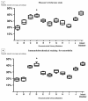
Fig 7. Percentage of the reconstructed bladder wall covered with smooth muscle.
Staining with (A) TRI and (B) anti-smoothelin staining. The regenerated bladder wall with a statistically similar (TRI [p = 0.03] and anti-smoothelin staining [p = 0.35]) smooth muscle content compared to the bladder wall in the control group (*).
Fig 8
Fig 8. The mechanical evaluation of reconstructed bladder wall based on Young’s elastic modulus.
Young’s modulus of intact and reconstructed bladder walls were compared to the digitally estimated content of smooth muscle content based on TRI staining average. Additionally, to reflect changes in the remodeling of the augmented bladder wall, Young’s modulus of Am and PLCL are presented. The presented values of smooth muscle content was rounded up.

References

    1. Witjes JA, Compérat E, Cowan NC, De Santis M, Gakis G, Lebrét T, et al. Guidelines on muscle-invasive and metastatic bladder cancer. In: EAU Guidelines, edition presented at the 28th EAU Annual Congress, Milan 2013. ISBN 978-90-79754-71-7
    1. Ordorica R, Wiegand LR, Webster JC, Lockhart JL. Ureteral replacement and onlay repair with reconfigured intestinal segments. J Urol. 2014: 191 1301–6. 10.1016/j.juro.2013.11.027 - DOI - PubMed
    1. Levine LA, Strom KH, Lux MM. Buccal mucosa graft urethroplasty for anteriorurethral stricture repair: evaluation of the impact of stricture location and lichen sclerosus on surgical outcome. J Urol. 2007: 178 2011–5. - PubMed
    1. Adamowicz J, Kowalczyk T, Drewa T. Tissue engineering of urinary bladder–current state of art and future perspectives. Cent European J Urol. 2013: 66 202–6. 10.5173/ceju.2013.02.art23 - DOI - PMC - PubMed
    1. Atala A, Bauer SB, Soker S, Yoo JJ, Retik AB. Tissue-engineered autologous bladders for patients needing cystoplasty. Lancet 2006: 367 1241–6. - PubMed

Substances