Diagnostic accuracy of oblique chest radiograph for occult pneumothorax: comparison with ultrasonography
- PMID: 26766962
- PMCID: PMC4711032
- DOI: 10.1186/s13017-016-0061-x
Diagnostic accuracy of oblique chest radiograph for occult pneumothorax: comparison with ultrasonography
Abstract
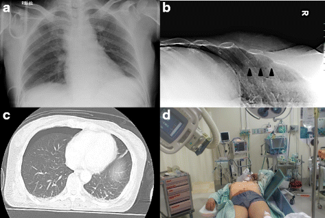
Backgraound: An occult pneumothorax is a pneumothorax that is not seen on a supine chest X-ray but is detected by computed tomography scanning. However, critical patients are difficult to transport to the computed tomography suite. We previously reported a method to detect occult pneumothorax using oblique chest radiography (OXR). Several authors have also reported that ultrasonography is an effective technique for detecting occult pneumothorax. The aim of this study was to evaluate the usefulness of OXR in the diagnosis of the occult pneumothorax and to compare OXR with ultrasonography.
Methods: All consecutive blunt chest trauma patients with clinically suspected pneumothorax on arrival at the emergency department were prospectively included at our tertiary-care center. The patients underwent OXR and ultrasonography, and underwent computed tomography scans as the gold standard. Occult pneumothorax size on computed tomography was classified as minuscule, anterior, or anterolateral.
Results: One hundred and fifty-nine patients were enrolled. Of the 70 occult pneumothoraces found in the 318 thoraces, 19 were minuscule, 32 were anterior, and 19 were anterolateral. The sensitivity and specificity of OXR for detecting occult pneumothorax was 61.4 % and 99.2 %, respectively. The sensitivity and specificity of lung ultrasonography was 62.9 % and 98.8 %, respectively. Among 27 occult pneumothoraces that could not be detected by OXR, 16 were minuscule and 21 could be conservatively managed without thoracostomy.
Conclusion: OXR appears to be as good method as lung ultrasonography in the detection of large occult pneumothorax. In trauma patients who are difficult to transfer to computed tomography scan, OXR may be effective at detecting occult pneumothorax with a risk of progression.
Keywords: Diagnosis; Lung ultrasound; Oblique chest radiograph; Occult pneumothorax.
Figures


References
-
- Surgeons ACo, editor. Advanced trauma life support course for doctors. Committee on Trauma. Instructors Course Manual.
LinkOut - more resources
Full Text Sources
Other Literature Sources
Medical

