. 2016 Jan 14:18:4.
doi: 10.1186/s12968-016-0223-8.

Microvascular ischemia in hypertrophic cardiomyopathy: new insights from high-resolution combined quantification of perfusion and late gadolinium enhancement

Affiliations

Microvascular ischemia in hypertrophic cardiomyopathy: new insights from high-resolution combined quantification of perfusion and late gadolinium enhancement

Adriana D M Villa et al. J Cardiovasc Magn Reson. .

Abstract

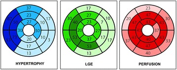
Background: Microvascular ischemia is one of the hallmarks of hypertrophic cardiomyopathy (HCM) and has been associated with poor outcome. However, myocardial fibrosis, seen on cardiovascular magnetic resonance (CMR) as late gadolinium enhancement (LGE), can be responsible for rest perfusion defects in up to 30% of patients with HCM, potentially leading to an overestimation of the ischemic burden. We investigated the effect of left ventricle (LV) scar on the total LV ischemic burden using novel high-resolution perfusion analysis techniques in conjunction with LGE quantification.

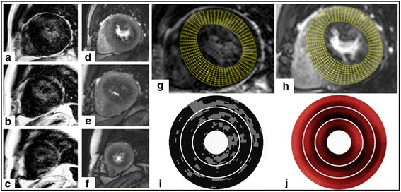
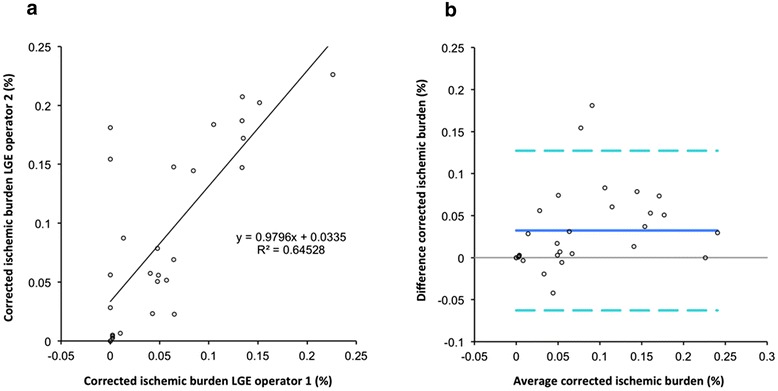
Methods: 30 patients with HCM and unobstructed epicardial coronary arteries underwent CMR with Fermi constrained quantitative perfusion analysis on segmental and high-resolution data. The latter were corrected for the presence of fibrosis on a pixel-by-pixel basis.

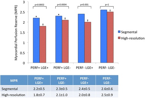
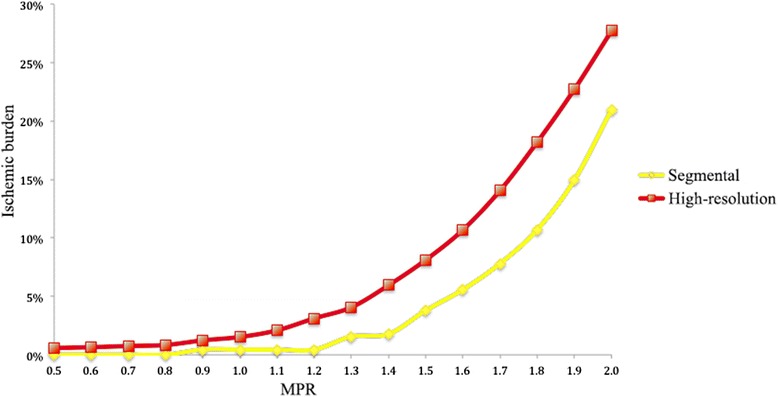
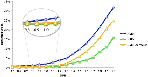
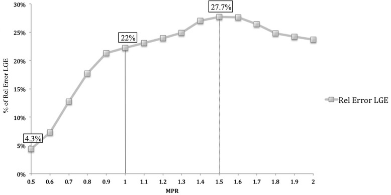
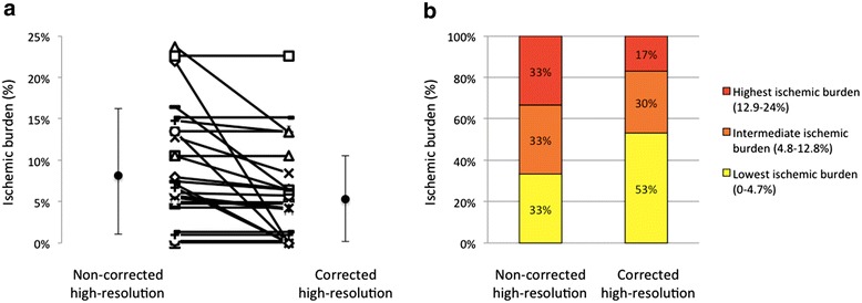
Results: High-resolution quantification proved more sensitive for the detection of microvascular ischemia in comparison to segmental analysis. Areas of LGE were associated with significant reduction of myocardial perfusion reserve (MPR) leading to an overestimation of the total ischemic burden on non-corrected perfusion maps. Using a threshold MPR of 1.5, the presence of LGE caused an overestimation of the ischemic burden of 28%. The ischemic burden was more severe in patients with fibrosis, also after correction of the perfusion maps, in keeping with more severe disease in this subgroup.

Conclusions: LGE is an important confounder in the assessment of the ischemic burden in patients with HCM. High-resolution quantitative analysis with LGE correction enables the independent evaluation of microvascular ischemia and fibrosis and should be used when evaluating patients with HCM.


Figures

Fig. 1
Fig. 1
Example of combined high-resolution fibrosis and perfusion mapping. a-c late gadolinium enhancement (LGE) images. d-f stress perfusion images. Top, middle and bottom rows correspond with basal, mid and apical slices respectively. Images (g) and (h) indicate high-resolution maps for LGE and stress perfusion respectively (basal slice only), with the grid used for high-resolution maps of LGE (I) and stress perfusion (J) shown below
Fig. 2
Fig. 2
Schematic representation of the distribution of hypertrophic segments, late gadolinium enhancement (LGE) and perfusion abnormalities based on visual assessment, expressed as percentages of the total cohort
Fig. 3
Fig. 3
Comparison between myocardial perfusion reserve (MPR) values obtained by using segmental and high-resolution quantification. Regions are classified according to the results of perfusion (PERF) and late gadolinium enhancement (LGE) visual assessment. *p < 0.0001 vs. segmental MPR of PERF-LGE- regions; † p < 0.0001 vs. all other groups for high-resolution MPR
Fig. 4
Fig. 4
Correlation between the different myocardial perfusion reserve (MPR) thresholds and the percentage of ischemic burden for segmental and high-resolution perfusion quantification
Fig. 5
Fig. 5
Correlation between the different myocardial perfusion reserve (MPR) thresholds and percentage of ischemic burden for high-resolution perfusion quantification of patients with a visual perfusion abnormality with and without including areas with overt late gadolinium enhancement (LGE), and in patients without LGE
Fig. 6
Fig. 6
Correlation between the different myocardial perfusion reserve (MPR) thresholds and relative error due to the inclusion of overt scar in the high-resolution perfusion analysis LGE: late gadolinium enhancement
Fig. 7
Fig. 7
a Non-corrected and corrected ischemic burden. Individual cases and average and standard deviation are shown. b Tertiles of non-corrected high-resolution ischemic burden and recategorization after correction for late gadolinium enhancement (LGE)
Fig. 8
Fig. 8
Impact of the inter-observer variability of late gadolinium enhancement (LGE) analysis on corrected ischemic burden measurements. a Pearson’s analysis. b Bland-Altman graph LGE: late gadolinium enhancement

References

    1. Elliott PM, Anastasakis A, Borger MA, Borggrefe M, Cecchi F, Charron P, et al. 2014 ESC Guidelines on diagnosis and management of hypertrophic cardiomyopathy. Eur Heart J. 2014;35:2733–79. doi: 10.1093/eurheartj/ehu199. - DOI - PubMed
    1. Marian AJ. On predictors of sudden cardiac death in hypertrophic cardiomyopathy*. J Am Coll Cardiol. 2003;41:994–6. doi: 10.1016/S0735-1097(02)03003-6. - DOI - PMC - PubMed
    1. Maron BJ, Shirani J, Poliac LC, Mathenge R, Roberts WC, Mueller FO. Sudden Death in Young Competitive Athletes: Clinical, Demographic, and Pathological Profiles. JAMA. 1996;276:199–204. doi: 10.1001/jama.1996.03540030033028. - DOI - PubMed
    1. McKenna W, Deanfield J, Faruqui A, England D, Oakley C, Goodwin J. Prognosis in hypertrophic cardiomyopathy: Role of age and clinical, electrocardiographic and hemodynamic features. Am J Cardiol. 1981;47:532–8. doi: 10.1016/0002-9149(81)90535-X. - DOI - PubMed
    1. McKenna WJ, England D, Doi YL, Deanfield JE, Oakley C, Goodwin JF. Arrhythmia in hypertrophic cardiomyopathy. I: Influence on prognosis. Br Heart J. 1981;46:168–72. doi: 10.1136/hrt.46.2.168. - DOI - PMC - PubMed

Publication types

MeSH terms