. 2016 Mar 15;310(6):G359-66.
doi: 10.1152/ajpgi.00388.2015. Epub 2016 Jan 14.

Predicting the activation states of the muscles governing upper esophageal sphincter relaxation and opening

Affiliations

Predicting the activation states of the muscles governing upper esophageal sphincter relaxation and opening

Taher I Omari et al. Am J Physiol Gastrointest Liver Physiol. .

Abstract

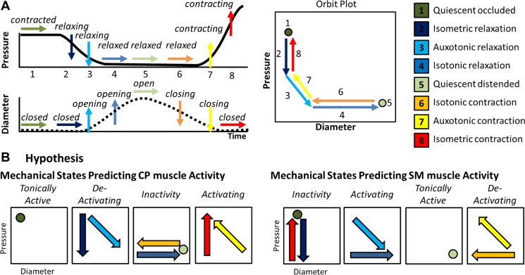
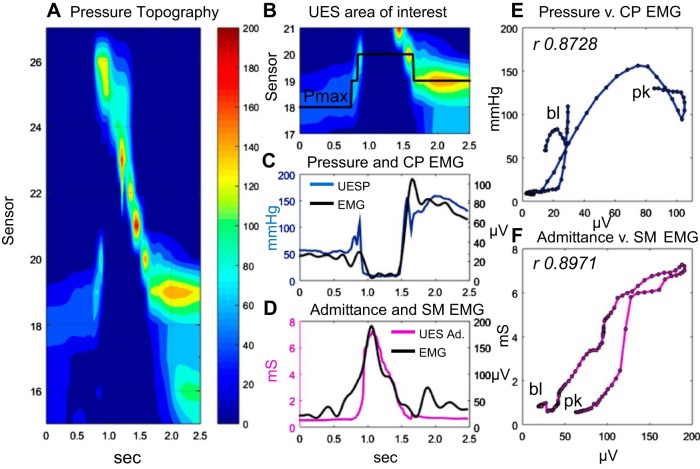
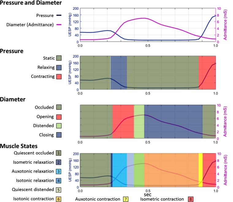
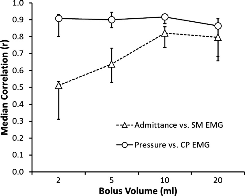
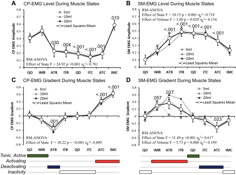
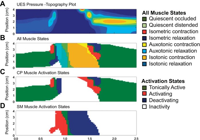
The swallowing muscles that influence upper esophageal sphincter (UES) opening are centrally controlled and modulated by sensory information. Activation and deactivation of neural inputs to these muscles, including the intrinsic cricopharyngeus (CP) and extrinsic submental (SM) muscles, results in their mechanical activation or deactivation, which changes the diameter of the lumen, alters the intraluminal pressure, and ultimately reduces or promotes flow of content. By measuring the changes in diameter, using intraluminal impedance, and the concurrent changes in intraluminal pressure, it is possible to determine when the muscles are passively or actively relaxing or contracting. From these "mechanical states" of the muscle, the neural inputs driving the specific motor behaviors of the UES can be inferred. In this study we compared predictions of UES mechanical states directly with the activity measured by electromyography (EMG). In eight subjects, pharyngeal pressure and impedance were recorded in parallel with CP- and SM-EMG activity. UES pressure and impedance swallow profiles correlated with the CP-EMG and SM-EMG recordings, respectively. Eight UES muscle states were determined by using the gradient of pressure and impedance with respect to time. Guided by the level and gradient change of EMG activity, mechanical states successfully predicted the activity of the CP muscle and SM muscle independently. Mechanical state predictions revealed patterns consistent with the known neural inputs activating the different muscles during swallowing. Derivation of "activation state" maps may allow better physiological and pathophysiological interpretations of UES function.

Keywords: cricopharyngeus muscle; deglutition; diameter; dysphagia; electromyography; impedance; neural pathways; pressure; submental muscles; upper esophageal sphincter.


Figures

Fig. 1.
Fig. 1.
Explanations and hypothesis in relation to upper esophageal sphincter (UES) mechanical states. A: typical pattern of pressure and diameter change that the UES undergoes during normal swallowing. A typical UES pressure profile is shown with arrows indicating periods when pressure is static (→), decreasing (↓), or increasing (↑). A typical diameter profile is shown with arrows indicating periods when the diameter is static (→), increasing (↑), or decreasing (↓). When the pressure and diameter data are interpreted together, different mechanical states can be defined based on the direction of pressure change and in relation to whether the lumen is open, closed, or changing in diameter. The relationship of diameter vs. pressure over time can also be visualized by way of an “orbit” plot. The mechanical states numbered 1–8 typify the normal sequence of UES contractility and UES opening during swallowing. B: we hypothesized that the 8 states can be consolidated into 4 groups defining when the muscle is tonically active, activating, deactivating, or inactive. Since the cricopharyngeus (CP) and extrinsic submental (SM) muscles perform mechanically reciprocal functions when activated, the association of states to muscle activity needs to be done separately for each muscle group type, depicted here as orbit plots.
Fig. 2.
Fig. 2.
An example of a 10-ml liquid swallow showing the relationship of CP-electromyography (EMG) to UES pressure and SM-EMG to UES intraluminal admittance. A: pressure topography plot of the entire pharyngoesophageal segment. B: area of interest defined for the UES high-pressure zone. Pmax, location of maximum axial UES pressure during the swallow. C: plot of UES pressure (defined by Pmax) and simultaneously recorded CP-EMG. D: plot of UES admittance (Ad.) (at Pmax) and simultaneously recorded SM-EMG. E: time correlation (Pearson rho) of CP-EMG vs. pressure data is shown for the period preswallow baseline (bl) to postrelaxation peak (pk) when diameter and pressure changes predominantly occur. F: time correlation of SM-EMG vs. admittance for the same baseline-to-peak period.
Fig. 3.
Fig. 3.
UES mechanical states determined from the UES pressure (UESP) and UES admittance profile from preswallow baseline to postrelaxation peak. Using the UES admittance and pressure data array for each of the swallows, we determined muscle states based on the direction of contraction or relaxation and in relation to the occluded or distended state of the lumen. Example is from the same swallow shown in Fig. 2.
Fig. 4.
Fig. 4.
Pearson rho time-correlations of SM-EMG vs. admittance and CP-EMG vs. pressure in relation to different volumes. The median of all subjects and interquartile ranges for each bolus volume are shown.
Fig. 5.
Fig. 5.
Normalized CP-EMG and SM-EMG activity recorded when different UES muscle states were predicted. A and B: EMG level, where higher values represent greater activity. C and D: EMG gradient, where positive/negative values indicate that EMG activity is increasing/decreasing with respect to time. Based on the EMG changes in these data with respect to mechanical state, conclusions were drawn with respect to the overall state of muscle activity present when the different muscle states were predicted. Below the graphs, the 8 states are consolidated into 4 groups defining when the muscle is tonically active (green), activating (red), deactivating (blue), or inactive (white). Graphs show the individual means for each state based on 5-, 10-, and 20-ml volumes and the overall estimated marginal mean for each state for all volumes combined (white circles with 95% confidence intervals). Repeated-measures (RM) ANOVA descriptive parameters are shown for each overall estimated marginal mean comparison. P values indicate pairwise significance vs. quiescent occluded (QO) state (post hoc test following Bonferroni correction). IMR, isometric relaxation; ATR, auxotonic relaxation; ITR, isotonic relaxation; QD, quiescent distended; ITC, isotonic contraction; ATC, auxotonic contraction; IMC, isometric contraction.
Fig. 6.
Fig. 6.
Generation of seamless maps of the spatiotemporal distribution of mechanical states (10-ml liquid swallow, previously shown in Figs. 2 and 3). A: pressure topography plot of the UES area of interest. B: muscle state map showing the appearance of 8 main muscle states over time. C: simplified muscle state map showing predicted CP muscle activity with states consolidated into 4 groups. D: simplified muscle state map showing predicted SM muscle activity with states consolidated into 4 groups.

References

    1. Asoh R, Goyal RK. Manometry and electromyography of the upper esophageal sphincter in the opossum. Gastroenterology 74: 514–520, 1978. - PubMed
    1. Clary MS, Daniero JJ, Keith SW, Boon MS, Spiegel JR. Efficacy of large-diameter dilatation in cricopharyngeal dysfunction. Laryngoscope 121: 2521–2525, 2011. - PubMed
    1. Cook IJ, Dodds WJ, Dantas RO, Massey B, Kern MK, Lang IM, Brasseur JG, Hogan WJ. Opening mechanisms of the human upper esophageal sphincter. Am J Physiol Gastrointest Liver Physiol 257: G748–G759, 1989. - PubMed
    1. Costa M, Wiklendt L, Arkwright JW, Spencer NJ, Omari T, Brookes SJH, Dinning PG. An experimental method to identify neurogenic and myogenic active mechanical states of intestinal motility. Front Syst Neurosci 7: 7, 2013. - PMC - PubMed
    1. Crary MA, Carnaby GD, Groher ME. Biomechanical correlates of surface electromyography signals obtained during swallowing by healthy adults. J Speech Lang Hear Res 49: 186–193, 2006. - PubMed

Publication types

LinkOut - more resources