. 2016 Mar 1;109(3):419-30.
doi: 10.1093/cvr/cvw005. Epub 2016 Jan 13.

Genetic lineage tracing discloses arteriogenesis as the main mechanism for collateral growth in the mouse heart

Affiliations

Genetic lineage tracing discloses arteriogenesis as the main mechanism for collateral growth in the mouse heart

Lingjuan He et al. Cardiovasc Res. .

Abstract

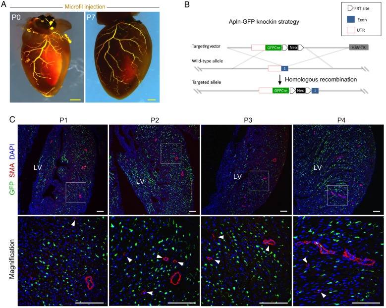
Aims: Capillary and arterial endothelial cells share many common molecular markers in both the neonatal and adult hearts. Herein, we aim to establish a genetic tool that distinguishes these two types of vessels in order to determine the cellular mechanism underlying collateral artery formation.

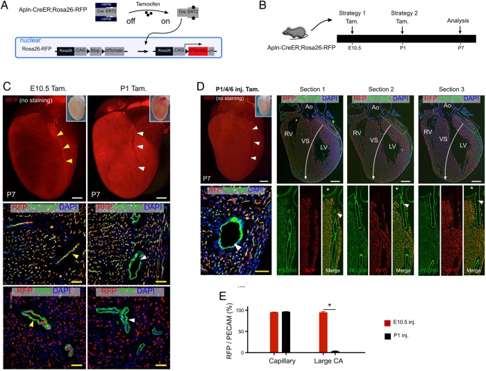
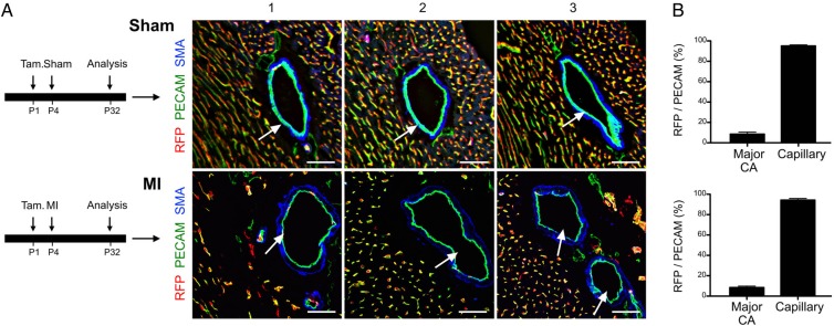
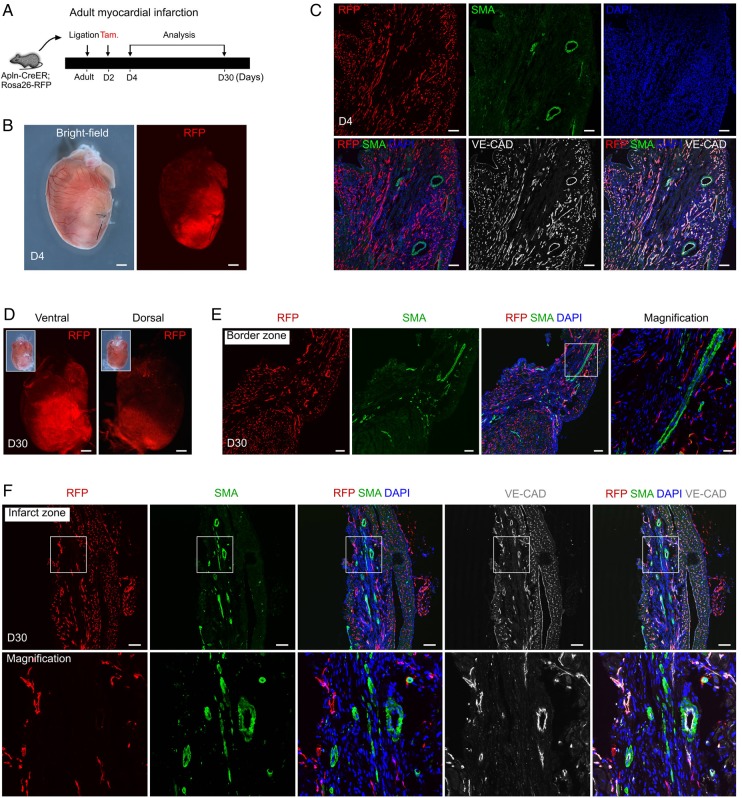
Methods and results: Using Apln-GFP and Apln-LacZ reporter mice, we demonstrate that APLN expression is enriched in coronary vascular endothelial cells. However, APLN expression is reduced in coronary arterial endothelial cells. Genetic lineage tracing, using an Apln-CreER mouse line, robustly labelled capillary endothelial cells, but not arterial endothelial cells. We leveraged this differential activity of Apln-CreER to study collateral artery formation following myocardial infarction (MI). In a neonatal heart MI model, we found that Apln-CreER-labelled capillary endothelial cells do not contribute to the large collateral arteries. Instead, these large collateral arteries mainly arise from pre-existing, infrequently labelled coronary arteries, indicative of arteriogenesis. Furthermore, in an adult heart MI model, Apln-CreER activity also distinguishes large and small diameter arteries from capillaries. Lineage tracing in this setting demonstrated that most large and small coronary arteries in the infarcted myocardium and border region are derived not from capillaries, but from pre-existing arteries.

Conclusion: Apln-CreER-mediated lineage tracing distinguishes capillaries from large arteries, in both the neonatal and adult hearts. Through genetic fate mapping, we demonstrate that pre-existing arteries, but not capillaries, extensively contribute to collateral artery formation following myocardial injury. These results suggest that arteriogenesis is the major mechanism underlying collateral vessel formation.

Keywords: Apln-CreER; Arterialization; Arteriogenesis; Collateral artery; Lineage tracing.


Figures

Figure 1
Figure 1
APLN expression in neonatal hearts. (A) Microfil injection into the coronary vascular circulation at P0 and P7 highlights the established, patent coronary vasculature. (B) A schematic model shows the knock-in strategy for generating the Apln-GFP allele by homologous recombination. (C) APLN expression was studied by analysis of post-natal day 1 (P1)–P4 neonatal hearts. Immunostaining for GFP as surrogate of APLN, SMA, and DAPI on sections of embryonic or neonatal Apln-GFP hearts revealed that Apln is expressed in capillaries, but is significantly reduced in large and small size arteries. Boxed regions are ventricle free wall and are magnified in figures in the lower panels. Arrowheads indicate APLNSMA+ coronary arteries. Each image is a representative of three to four individual samples. Scale bars: yellow, 0.5 mm; white, 100 µm.
Figure 2
Figure 2
Labelling of coronary capillaries and arteries by Apln-CreER. (A) A schematic figure showing tamoxifen induction of CreER activity and genetic labelling of Apln+ cells. (B) Strategies for tamoxifen induction (Tam.) and analysis of coronary artery labelling in Apln-CreER; Rosa26-RFP mice. (C) Whole-mount view (top) and stained sections (below) of P7 Apln-CreER; Rosa26-RFP hearts following tamoxifen treatment. Tamoxifen induction at E10.5, but not P1, labels endothelial cells (PECAM+) of major coronary arteries (yellow vs. white arrowheads). (D) Whole-mount view and serial sections (50 µm apart, sections progress left to right from ventral to dorsal panels) of P7 Apln-CreER; Rosa26-RFP hearts following serial neonatal tamoxifen induction (P1/4/6). The endothelium of the coronary arteries is sparsely labelled (white arrowheads). Asterisks indicate orifices in the aorta. (E) Quantification of RFP+PECAM+ cells of the capillaries and large coronary arteries in the outer myocardial wall of P7 hearts following tamoxifen induction at E10.5 or P1. *P < 0.05, n = 5–6 per group. LV, left ventricle; RV, right ventricle; VS, ventricular septum; Ao, aorta. Scale bars: white bar, 0.5 mm; yellow bar, 50 µm.
Figure 3
Figure 3
Establishment of a neonatal MI injury model. (A) TTC (2,3,5-triphenyltetrazolium chloride) staining of P4 hearts demarcates damaged myocardium (infarct zone) after ligation of the LAD coronary artery (black arrow). (B) Microfil-injected heart 28 days (28d) after sham surgery. White arrowhead points to the non-ligated coronary artery. (C and D) Ventral and left-sided views of microfil-injected heart 28 days after MI. White arrowheads indicate the ligation site of the previous LAD coronary artery. Green arrows indicate newly established major coronary arteries that bypass the ligation point. n = 3–4. Scale bars: 1 mm. (E) Functional measurement of MI hearts by M-mode echocardiography showing systolic and diastolic cardiac contractions. Two indicators of cardiac output, EF and FS, calculated from the echocardiography, were markedly reduced in MI hearts when compared with sham-operated controls. n = 4. (F) Masson's trichrome and Sirius Red staining of MI and control hearts. MI was performed on P4 neonatal hearts by ligation of the LAD artery, and hearts were collected 28 days after MI (at P32). Significant fibrosis was induced in the left apex of the ventricle, indicating effective MI induction. All the mice used here are wild-type ICR/C57BL6/J-mixed background mice. n = 4. Black bars, 1 mm.
Figure 4
Figure 4
Fate map of arteries and capillaries following MI. (A) The experimental timeline for tamoxifen induction of Apln-CreER activity in the post-natal heart after sham or surgery. Immunostaining for RFP, PECAM, and SMA on sections of P32 Apln-CreER; Rosa26-RFP hearts after tamoxifen treatment (Tam.) at P1. White arrows point to the collateral arteries, which were not significantly labelled in either sham or MI hearts, suggesting that these major large diameter adult coronary arteries are not formed de novo from labelled capillaries at P1. (B) The extent of labelling is quantified as the percentage of RFP+ endothelial cells over total PECAM+ endothelial cells. There is significant difference between major coronary artery and capillaries in RFP+ cell percentage (P < 0.05, n = 3–4). Scale bars: 50 µm.
Figure 5
Figure 5
Robust labelling of large coronary arteries following E10.5 tamoxifen treatment. (A) Immunostaining for RFP and PECAM on sections from Apln-CreER; Rosa26-RFP hearts showed robust labelling of arterial endothelial cells (RFP+, white arrows), as well as capillaries. Epi, epicardium; endo, endocardium. (B) The experimental strategy is shown above. Below, quantification of the labelling efficiency is shown. Labelling efficiency was calculated as the percentage of RFP+ cells in PECAM+ vessels of the outer myocardial wall. Scale bars, 100 µm. (C) Immunostaining for RFP and PECAM showed that collateral arterial endothelial cells (white arrows) were labelled in the MI hearts. Tamoxifen was administered at E10.5. Scale bars, 50 µm. (D) Quantification of the percentage of RFP+ endothelial cells. Each image is representative of three individual samples.
Figure 6
Figure 6
Labelling of small coronary arteries in the neonatal and adult heart. (A) A schematic figure shows the strategy for tamoxifen induction (Tam) and analysis (at P4 and P32). (B) Immunostaining for RFP, SMA, and PECAM on sections of P4 Apln-CreER; Rosa26-RFP hearts after Tamoxifen treatment at P1. Arrowheads indicate a small size artery (<30 µm); asterisks indicate large artery (>60 µm); Endo, endocardium. Scale bars, 100 µm. (C) Quantification of labelling efficiency, defined as the percentage of RFP+PECAM+ endothelial cells compared with total PECAM+ vascular endothelial cells. Student's t-test was used to analyse differences, and values are shown as mean ± SEM; *P < 0.05; n = 3–4.
Figure 7
Figure 7
Coronary arteries in the infarcted myocardium are derived from pre-existing arteries but not from capillaries. (A) Schematic showing the experimental strategy for injury, tamoxifen induction, and analysis. (B) Whole-mount bright-field and fluorescent views of Apln-CreER; Rosa26-RFP hearts at day 4 (D4) after MI. (C) Immunostaining for RFP, SMA, and VE-CAD showed that Apln-CreER robustly labels VE-CAD+SMA coronary capillaries, but not VE-CAD+SMA+ coronary arteries. (D) Whole-mount view of Apln-CreER; Rosa26-RFP hearts at D30 after MI. Insets are bright-field views of hearts. (E) Immunostaining for RFP and SMA showed that Apln-CreER-labelled capillaries do not form coronary arteries in the border zone. (F) Immunostaining for RFP, SMA, and VE-CAD showed that Apln-CreER-labelled coronary capillaries do not contribute significantly to coronary arteries in the infarct zone. Scale bars: 1 mm in (B) and (D) and 100 µm in (C), (E), and (F). Each image is representative of three individual samples.

References

    1. Helisch A, Schaper W. Arteriogenesis: the development and growth of collateral arteries. Microcirculation 2003;10:83–97. - PubMed
    1. Rubanyi GM. Mechanistic, technical, and clinical perspectives in therapeutic stimulation of coronary collateral development by angiogenic growth factors. Mol Ther 2013;21:725–738. - PMC - PubMed
    1. Ito W, Arras M, Winkler B, Scholz D, Schaper J, Schaper W. Monocyte chemotactic protein-1 increases collateral and peripheral conductance after femoral artery occlusion. Circ Res 1997;80:829–837. - PubMed
    1. Arras M, Ito W, Scholz D, Winkler B, Schaper J, Schaper W. Monocyte activation in angiogenesis and collateral growth in the rabbit hindlimb. J Clin Invest 1998;101:40–50. - PMC - PubMed
    1. Belmadani S, Matrougui K, Kolz C, Pung YF, Palen D, Prockop DJ, Chilian WM. Amplification of coronary arteriogenic capacity of multipotent stromal cells by epidermal growth factor. Arterioscler Thromb Vasc Biol 2009;29:802–808. - PMC - PubMed

Publication types

MeSH terms