. 2015 Dec 22;11(48):6458-66.
doi: 10.1002/smll.201502972. Epub 2015 Nov 19.

Bioorthogonal Click Chemistry-Based Synthetic Cell Glue

Affiliations

Bioorthogonal Click Chemistry-Based Synthetic Cell Glue

Heebeom Koo et al. Small. .

Abstract

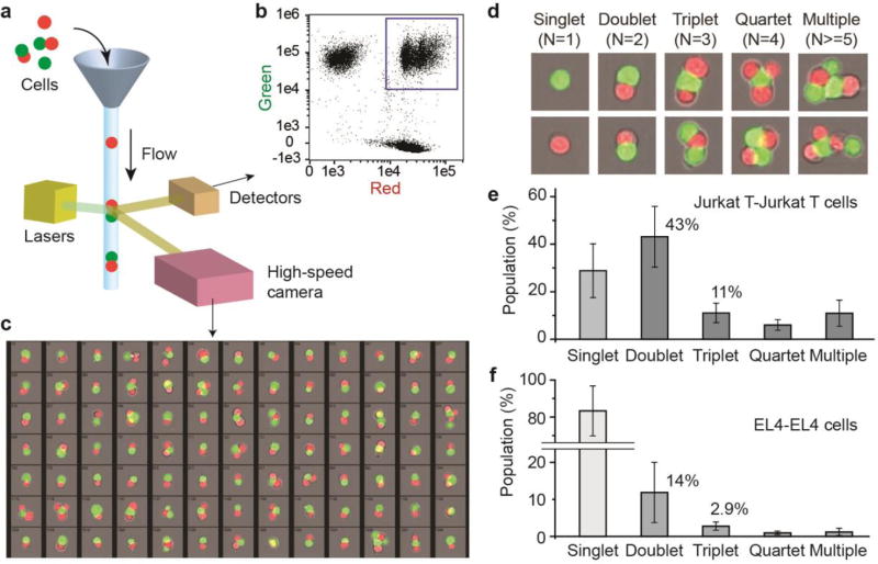
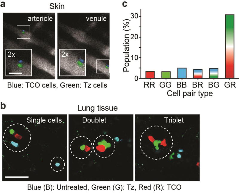
Artificial methods of cell adhesion can be effective in building functional cell complexes in vitro, but methods for in vivo use are currently lacking. Here, a chemical cell glue based on bioorthogonal click chemistry with high stability and robustness is introduced. Tetrazine (Tz) and trans-cyclooctene (TCO) conjugated to the cell surface form covalent bonds between cells within 10 min in aqueous conditions. Glued, homogeneous, or heterogeneous cell pairs remain viable and stably attached in a microfluidic flow channel at a shear stress of 20 dyn cm(-2) . Upon intravenous injection of assembled Jurkat T cells into live mice, fluorescence microscopy shows the trafficking of cell pairs in circulation and their infiltration into lung tissues. These results demonstrate the promising potential of chemically glued cell pairs for various applications ranging from delivering therapeutic cells to studying cell-cell interactions in vivo.

Keywords: cell adhesion; cell delivery; click chemistry; metabolic glycoengineering; tissue engineering.


Figures

Figure 1
Figure 1
Analysis of viability and function of glued cells. (a) Measured amount of Tz and TCO groups on cell surface after chemical modification for four different cell lines. Error bars, s.d.; *, t-test P < 0.005 (sample n=10). (b) Illustration of cellular gluing between suspension (red) and adhesion (green) cells. (c) Fluorescence images of the glued cells in a microfluidic chamber after washing with a flow at 1 ml/mm. Scale bar, 50 µm (d) Viability of Jurkat-Jurkat glued cells measured using calcein AM/Ethidium homodimer 1 assay after incubation for 1 day in culture. (e) IL-2 secretion from glued Jurkat T cells. Error bars, s.d. (sample n=5). (f) Microscopic images showing the migration of NIH3T3 cells (green) carrying Jurkat T cells (red) glued on their surface. Scale bar, 100 µm.
Figure 2
Figure 2
Analysis of the binding force between glued cells. (a) Illustration of the flowing test on the glued cells in microfluidic chambers. (b) Fluorescence images of the glued Jurkat T cells on A549 cells. Scale bar, 50 µm. Flow at 60 ml/min was applied for duration of 1 min, at 10 min (Day 0) and 24 hours (Day 1) after Tz-TCO gluing. The ratio of cell numbers in the glue pairs after flow for Jurkat-A549 (d) and Jurkat-NIH3T3 (e) cells.
Figure 3
Figure 3
Microscopy and cytometry analysis. (a) Fluorescence images of glued Jurkat T cells in suspension. Scale bar, 100 µm. (b) High-resolution image of two-cell doublet and four-cell quartet glued cells (circles). Scale bar, 50 µm. (c) SEM images of doublet (top) and triplet (bottom) glued Jurkat T cells (pseudo colored green and red). Scale bar, 5 µm. Flow cytometry data of Jurkat T cells glued with different concentrations of Tz and TCO (d) or by azide-DBCO click chemistry (e). Inset, a schematic of DBCO-PEG4-DBCO.
Figure 4
Figure 4
Analysis of glued cells by imaging cytometry. (a) Illustration showing simultaneous flow cytometry and microscopic imaging. (b) Measured cytometry graph. The population in the box corresponds to glued cells. (c) Cytometry images showing glued population of green-and red-labeled Jurkat T cells after focus filtering. (d) Representative captured images of various cell groups. (e) The measured statistics of various cell groups for the case of Jurkat-Jurkat cells. (f) Statistics for EL4-EL4 cells.
Figure 5
Figure 5
Cells injected into mice. (a) Intravital images of glued Jurkat T cells flowing in the blood vessels of the ear skin. Scale bar, 100 µm. (b) Fluorescence images of Jurkat T cells in lung tissues harvested about 20 min after injection. Scale bar, 100 µm. (c) Measured population of cells in contact in the lung tissues. R, G, and B represent the color of cell: ‘GR’ indicates cell pairs consisting of Tz-modified (green) and TCO-modified (red) cells.
Scheme 1
Scheme 1
Illustration of the cellular gluing method based on metabolic glycoengineering and double click chemistry. (Chemical structure was corrected)

References

    1. Ahrends R, Ota A, Kovary KM, Kudo T, Park BO, Teruel MN. Science. 2014;344:1384–1389. - PMC - PubMed
    2. Meshel AS, Wei Q, Adelstein RS, Sheetz MP. Nat Cell Biol. 2005;7:157–164. - PubMed
    1. Lee S, Mandic J, Van Vliet KJ. Proceedings of the National Academy of Sciences. 2007;104:9609–9614. - PMC - PubMed
    2. Aceto N, Bardia A, Miyamoto DT, Donaldson MC, Wittner BS, Spencer JA, Yu M, Pely A, Engstrom A, Zhu HL, Brannigan BW, Kapur R, Stott SL, Shioda T, Ramaswamy S, Ting DT, Lin CP, Toner M, Haber DA, Maheswaran S. Cell. 2014;158:1110–1122. - PMC - PubMed
    1. Ohashi K, Yokoyama T, Yamato M, Kuge H, Kanehiro H, Tsutsumi M, Amanuma T, Iwata H, Yang J, Okano T, Nakajima Y. Nat. Med. 2007;13:880–885. - PubMed
    2. Karp JM, Leng Teo GS. Cell stem cell. 2009;4:206–216. - PubMed
    1. Gartner ZJ, Bertozzi CR. Proceedings of the National Academy of Sciences. 2009;106:4606–4610. - PMC - PubMed
    2. Todhunter ME, Jee NY, Hughes AJ, Coyle MC, Cerchiari A, Farlow J, Garbe JC, LaBarge MA, Desai TA, Gartner ZJ. Nat Meth. 2015 doi: 10.1038/nmeth.3553. in press. - DOI - PMC - PubMed
    1. Vogel K, Glettenberg M, Schroeder H, Niemeyer CM. Small. 2013;9:255–262. - PubMed

Publication types