. 2016 Jan 14;5(1):e002484.
doi: 10.1161/JAHA.115.002484.

Full Expression of Cardiomyopathy Is Partly Dependent on B-Cells: A Pathway That Involves Cytokine Activation, Immunoglobulin Deposition, and Activation of Apoptosis

Affiliations

Full Expression of Cardiomyopathy Is Partly Dependent on B-Cells: A Pathway That Involves Cytokine Activation, Immunoglobulin Deposition, and Activation of Apoptosis

Andrea M Cordero-Reyes et al. J Am Heart Assoc. .

Abstract

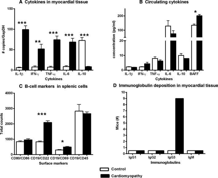
Background: Limited information exists on the role of B-cell-dependent mechanisms in the progression of heart failure (HF). However, in failing human myocardium, there is evidence of deposition of activated complement components as well as anticardiac antibodies. We aimed to determine the contribution of B-cells in HF progression using a nonsurgical mouse model of nonischemic cardiomyopathy (CMP).

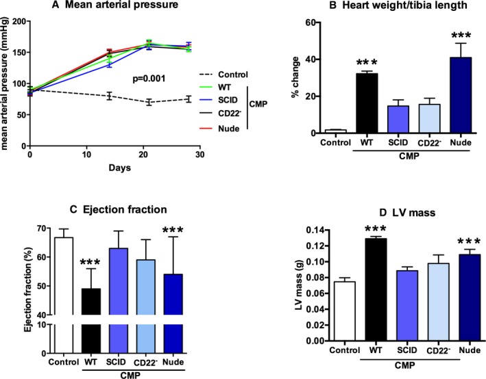
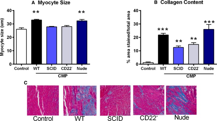
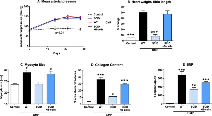
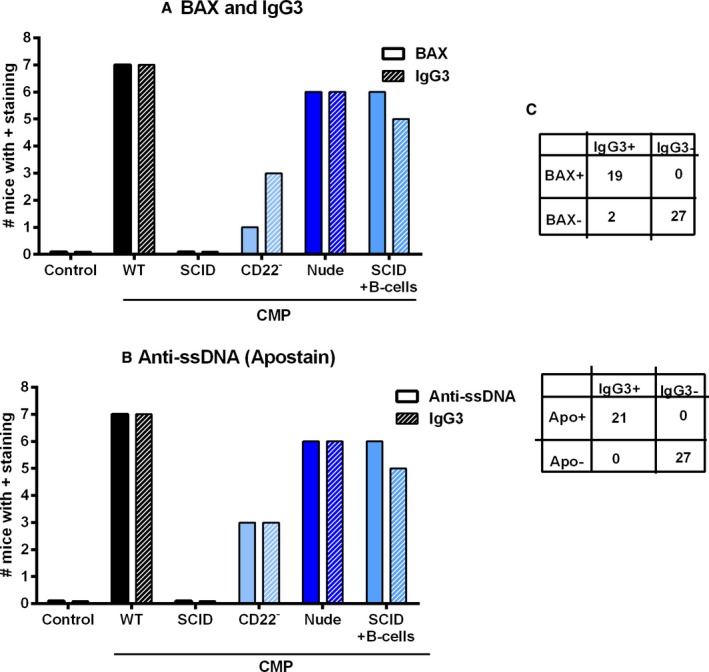
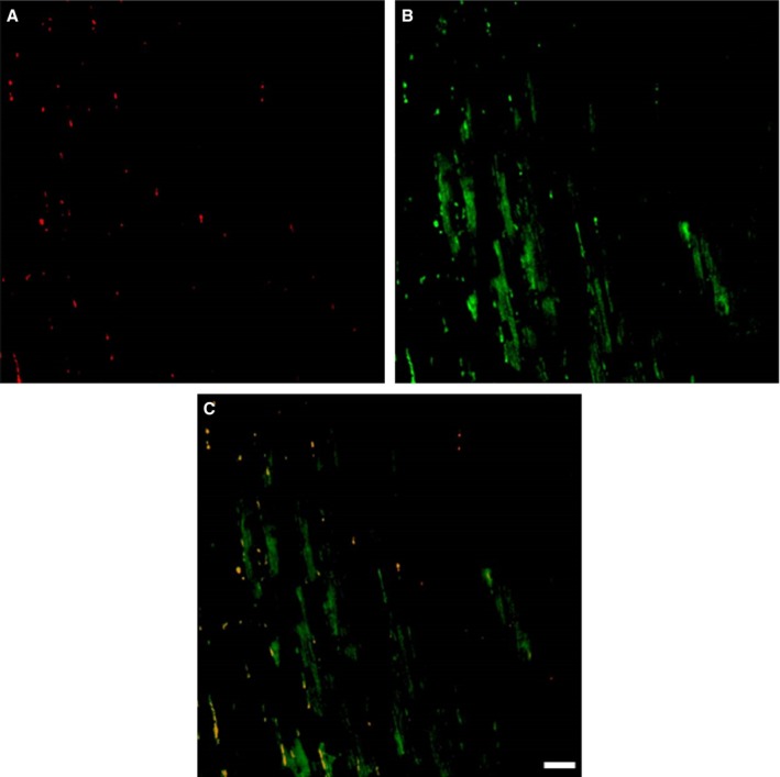
Methods and results: CMP protocol involved the use of l-NAME and NaCl in the drinking water and angiotensin-II infusion for 35 days. At day 35, mice were analyzed by cardiac magnetic resonance imaging, gene expression, and histology. Mice (12 weeks old) were divided into 4 groups, all in C57BL/6 background: wild-type (WT) CMP; severe combined immunodeficiency (SCID) CMP (T- and B-cell deficient); CD22(-) CMP (B-cell depleted); and Nude CMP (T-cell deficient), with their respective controls. We performed B-cell depletion and reconstitution protocols. The protective effect of B-cell depletion was demonstrated by a significant reduction of cell hypertrophy and collagen deposition and a preserved ejection fraction in the CD22(-) CMP group compared to WT CMP. Once SCID mice underwent B-cell reconstitution with isolated CMP B-cells, the CMP phenotype was restored. Furthermore, deposition of IgG3 and apoptosis in the myocardium follows the development of CMP; in addition, in vitro studies demonstrated that activated B-cells stimulate collagen production by cardiac fibroblasts.

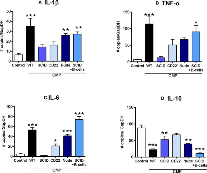
Conclusions: The absence of B-cells in this model of HF resulted in less hypertrophy and collagen deposition, preservation of left ventricular function, and, in association with these changes, a reduction in expression of proinflammatory cytokines, immunoglobulin G deposition, and apoptosis in the myocardium. Taken together, these data suggest that B-cells play a contributory role in an angiotensin-II-induced HF model.

Keywords: antibodies; cardiomyopathy; immune system; lymphocytes; remodeling.


Figures

Figure 1
Figure 1
Immune system changes in a mouse model of nonischemic cardiomyopathy. A, Proinflammatory cytokines are upregulated in the myocardial tissue milieu, whereas (B) cytokines in the circulation only show a significant increase in BAFF expression. C, Surface marker profiles of splenic B‐cells of CMP mice compared to controls show an overall increase in activated B‐cells (CD19/CD69), but not total B‐cell numbers, and/or total leucocytes (CD19/CD45) side‐scatter horizontal was used for B‐cell markers (CD19) and side‐scatter vertical was used for other markers (CD86, CD69, and CD45). D, Immunofluorescence assays determined the deposition of immunoglobulins in myocardial tissue: only IgG3 was present and distributed along the sarcolemma. All P values compare controls versus all other groups: *P<0.05; **P<0.01; ***P<0.001. BAFF indicates B‐cell activating factor; CMP indicates cardiomyopathy; IFN‐γ, interferon‐gamma; Ig, immunoglobulin; IL, interleukin; TNF‐α, tumor necrosis factor alpha.
Figure 2
Figure 2
B‐cells are not required for hypertension, but for cardiomyopathy. Shown is the hemodynamic response of untreated mice and cardiomyopathy treated mice in the following groups: wild type (WT CMP); mice that lack B‐ and T‐cells (SCID CMP); mice treated with an antibody to eliminate CD22+ cells (CD22 CMP); or mice that lack T‐cells (Nude CMP). A, Hypertensive response was equal among all the CMP‐treated groups, independent of B‐ and T‐cell compartments, compared to controls. B, As shown by the percent change in heart weight per tibia length, greater increases were observed in the WT CMP and Nude CMP groups compared to the SCID CMP and CD22 CMP groups. C, LVEF, as measured by cMRI at the end of the cardiomyopathy protocol, is significantly reduced in WT CMP and Nude CMP mice compared to controls. The SCID CMP and CD22 CMP groups have a significantly higher LVEF than the WT CMP mice. D, Changes in LV mass followed an inverse pattern to the changes in LVEF, with LV mass elevated in WT CMP compared to controls (n=15 per group). All P values compare controls versus all other groups: *P<0.05; **P<0.01; ***P<0.001.
Figure 3
Figure 3
Lack of B‐cells attenuates adverse remodeling in cardiomyopathy. A, The greatest increase in myocyte size compared to untreated controls was observed in the WT CMP and Nude CMP groups, whereas in mice of the CD22 CMP and SCID CMP groups, the increase in myocyte size was insignificant compared to controls. B, A significant increase in collagen content was observed in WT CMP and Nude CMP mice compared to controls, whereas only a modest increase in collagen content was observed in the SCID CMP and CD22 CMP groups (n=15 per group). C, Representative pictures of myocardial tissue sections stained for trichrome in all 5 groups. All P values compare controls versus all other groups: *P<0.05; **P<0.01; ***P<0.001.
Figure 4
Figure 4
Reconstitution of B‐cells in SCID mice restores the heart failure phenotype. The figure shows untreated controls and mice treated to produce cardiomyopathy in the following backgrounds: WT; SCID; and SCID with B‐cell reconstitution (SCID+B‐cells). Once we reconstituted B‐cells in SCID mice and induced CMP, (A) hypertensive response was similar, (C) myocyte size was increased, and this effect was also observed with the (B) percent change in heart weight to tibia length and (D) collagen content. E, BNP expression was high in WT CMP group, lower in B‐cell‐deficient mice (CD22 CMP [not shown] or SCID CMP), and elevated in SCID mice with B‐cell reconstitution (n=15 per group). All P values compare controls versus all other groups: *P<0.05; **P<0.01; ***P<0.001. BNP indicates brain natriuretic peptide; CMP, cardiomyopathy; SCID, severe combined immune deficiency; WT, wild type.
Figure 5
Figure 5
Inflammatory cytokine profile in B‐cell‐deficient mice and B‐cell‐reconstituted mice compared to controls with CMP. Quantitative gene expression of inflammatory markers in cardiac tissues at the end of the treatment protocol, the WT CMP group was characterized by increase in (A) IL‐1β, (B) TNF‐α, and (C) IL‐6 expression compared to untreated controls. In the groups with B‐cell‐deficient mice (SCID CMP, CD22 CMP, and Nude CMP), there was reduced expression of these genes. After B‐cell reconstitution in the SCID CMP group, expression of TNF‐α, IL‐6, and IL‐1β increased. D, IL‐10 expression was significantly reduced in the WT CMP group and increased to levels comparable to the untreated controls in the SCID CMP and CD22 CMP groups. All P values compare controls versus all other groups: *P<0.05; **P<0.01; ***P<0.001. CMP indicates cardiomyopathy; IL, interleukin; SCID, severe combined immune deficiency; TNF‐α, tumor necrosis factor alpha; WT, wild type.
Figure 6
Figure 6
Markers of antibody deposition and activation of apoptosis in myocardial tissue of control and CMP mice. A, IgG3 expression was present in most mice in the WT CMP group. Fewer mice in the B‐cell‐deficient groups, SCID CMP and CD22 CMP, were positive for IgG3. In contrast, higher numbers of mice in the T‐cell‐depleted but with intact B‐cell function groups, Nude CMP or SCID with B‐cell reconstitution CMP, were positive for IgG3. Bax expression in myocardium of control mice and various CMP‐treated groups is also shown, with a pattern of expression similar to IgG3. C, The 2×2 tables show the distribution of IgG3 and Bax or IgG3 and anti‐ssDNA staining in all the pooled analyzed myocardial tissue samples (n=8/group). B. A similar pattern was observed when compared to Ant‐ssDNA staining.
Figure 7
Figure 7
Colocalization of IgG3 with markers of apoptosis. Figure shows representative images where BAX‐positive areas stained red (A) and IgG3‐positive areas stained green (B), with dual staining in the bottom panel in yellow (C). Scale bar, 100 μm. BAX indicates B‐cell lymphoma‐2‐associated X protein; Ig, immunoglobulin.
Figure 8
Figure 8
Markers of proliferation and collagen expression in isolated cardiac fibroblasts after stimulation with activated B‐cell products. Figure shows gene expression profile in cultured cardiac fibroblasts where there is increased expression of NF‐κB, PCNA, ki‐67, IL‐6, and collagen (Coll) I and III with either HF B‐cell supernatant or stimulated B‐cell (LPS and CpG‐ODN) supernatant. HF indicates heart failure; IL, interleukin; NF‐κB, nuclear factor kappa B; PCNA, proliferating cell nuclear antigen; RT‐PCR, reverse‐transcriptase polymerase chain reaction.

References

    1. Suzuki H, Sato R, Sato T, Shoji M, Iso Y, Kondo T, Shibata M, Koba S, Katagiri T. Time course of changes in the levels of interleukin 6 in acutely decompensated heart failure. Int J Cardiol. 2005;100:415–420. - PubMed
    1. Milani RV, Mehra MR, Endres S, Eigler A, Cooper ES, Lavie CJ Jr, Ventura HO. The clinical relevance of circulating tumor necrosis factor‐alpha in acute decompensated chronic heart failure without cachexia. Chest. 1996;110:992–995. - PubMed
    1. Peschel T, Schonauer M, Thiele H, Anker SD, Schuler G, Niebauer J. Invasive assessment of bacterial endotoxin and inflammatory cytokines in patients with acute heart failure. Eur J Heart Fail. 2003;5:609–614. - PubMed
    1. Sato Y, Takatsu Y, Kataoka K, Yamada T, Taniguchi R, Sasayama S, Matsumori A. Serial circulating concentrations of C‐reactive protein, interleukin (IL)‐4, and IL‐6 in patients with acute left heart decompensation. Clin Cardiol. 1999;22:811–813. - PMC - PubMed
    1. Torre‐Amione G, Orrego CM, Khalil N, Kottner‐Assad C, Leveque C, Celis R, Youker KA, Estep JD. Therapeutic plasma exchange a potential strategy for patients with advanced heart failure. J Clin Apher. 2010;25:323–330. - PubMed

Publication types

MeSH terms