. 2016 Mar 1;7(9):9815-31.
doi: 10.18632/oncotarget.6890.

Identification of stem-like cells and clinical significance of candidate stem cell markers in gastric cancer

Affiliations

Identification of stem-like cells and clinical significance of candidate stem cell markers in gastric cancer

Xiaowei Zhang et al. Oncotarget. .

Abstract

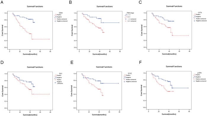
The existence of gastric cancer stem cells (CSCs) has not been definitively proven and specific cell surface markers for identifying gastric CSCs have largely not been identified. Our research aimed to isolate potential gastric CSCs and clarify their clinical significance, while defining markers for GCSC identification and verification. Here, we report that spheroid cells possess stem cell-like properties, and overexpress certain stem cell markers. CD133 or CD44-positive cells also exhibit properties of CSCs. The expression of Oct4, Sox2, Gli1, CD44, CD133, p-AKT, and p-ERK was significantly higher in metastatic lesions compared to that in primary lesions. Elevated expression of some of these proteins was correlated with a more aggressive phenotype and poorer prognosis, including Oct4, Sox2, Gli1, CD44, and p-ERK. Multivariate Cox proportional hazards model analysis showed that only CD44 is an independent factor. Knockdown of CD44 down-regulated the stem cell-like properties, which was accompanied by the down-regulation of p-ERK and Oct4. Oct4 overexpression could reverse the decreased CSCs properties induced by CD44 knockdown. Taken together, our research revealed that spheroid cell culture, and CD133 or CD44-labeled FACS methods can be used to isolate gastric CSCs. Some CSC markers have clinical significance in predicting the prognosis. CD44 is an independent prognostic factor and maintains the properties of CSCs in CD44-p-ERK-Oct4 positive feedback loop.

Keywords: CD133; CD44; cancer stem cell; gastric cancer; stem cell marker.


Conflict of interest statement

CONFLICT OF INTEREST

The authors declare no conflict of interest.

Figures

Figure 1
Figure 1. Sphere cells isolated from gastric cancer by serum free culture have the properties of CSCs
A. tumorigenic spheres are derived from gastric cancer cell lines or primary gastric cancer cells by serum free culture. B. isolated spheroid cells overexpress stem cell markers. The expression of stem cell markers in spheroid cells (SCs) from gastric cancer cell lines or primary gastric cancer cells and their parental cells (Con) was detected by Western blotting. C. spheroid cells or parental cells are treated with EPI in serum-free RPML-1640 culture medium at the indicated time points (n = 5). D. isolated spheroid cells overexpress chemo-resistance-related proteins MDR1 and ABCG2, as detected by Western blot. E. spheroid cells have higher tumorigenicity in vivo compared with parental cells. Pictures of xenografts after subcutaneous injection of control and sphere cells in SCID mice are depicted (n = 4). (data are represented as mean +/− SD).
Figure 2
Figure 2. CD44-positive cancer cells have properties of CSCs
A. CD44-negative (CD44-) and CD44-positive (CD44+) cells were sorted with a FACS Aria (BD Biosciences); CD44-positive cells account for 86% of the total cells. B. CD44-positive cells have higher tumorigenicity in vivo compared with CD44-negative cells (n = 5). C. CD44-positive cells display greater chemo-resistance. The CCK8 assay was used to evaluate the sensitivity of CD44-positive or CD44-negative cells to cytotoxic agent 5-Fu treatment. D. CD44-positive cells have a higher migration and invasion ability. Transwell migration or invasion assays were analyzed using the Corning chamber. The migration and invasion were photographed (upper panel) and quantified (lower panel), E. Representative H&E-stained sections of the lung tissues isolated from NOD-SCID mice that injected with CD44-negative or CD44-positive cells through the lateral tail vein. The numbers of metastases in the lungs were counted. F. Typical EMT morphology photos were shown (left panel); The expression of Oct4, p-ERK, CD44, E-cadherin, Vimentin proteins were detected by western blots analysis in CD44 negative or CD44 positive cells (right panel). (Data are represented as mean +/− SD)
Figure 3
Figure 3. CD133-positive cancer cells have the properties of CSCs
A. CD133-negative and CD133-positive cells were sorted with a FACS (BD Biosciences) method labeled by CD133 or CD44 antibody, and the percentage of CD133-positive cells in MKN45 cells was 0.78% of the total cells. B. CD133-positive cells display higher tumorigenicity in vivo compared with CD133-negative cells. Growth curves of tumors after subcutaneous injection of CD133-positive and CD133-negative cells in SCID mice are depicted. Data represent the mean ± SD (n = 3). C. CD133-positive cells are more chemo-resistance. CCK8 assay was used to evaluate the sensitivity of cytotoxic agent 5-Fu between CD133-positive and CD133-negative groups. D. CD133-positive cells has higher migration and invasion ability. Transwell migration or invasion assays were analyzed using the Corning chamber. The migration and invasion were photographed (upper panel) and quantified (lower panel), E. Typical EMT morphology photos were shown (left panel); The expression of CD133, E-cadherin, Vimentin proteins were detected by western blots in CD133 negative or CD133 positive cells (right panel). (Data are represented as mean +/− SD)
Figure 4
Figure 4. Representative figures of several CSC-related markers or proteins in, gastric tumors, its surrounding normal tissues and paired metastatic cancer samples
A. primary cancer tissues express less Sox2 compared with the paired metastatic cancer tissues. In panel B., primary cancer tissues express less Gli1 compared with metastatic cancer tissues. C. primary cancer tissues expresses less CD44 compared with metastatic cancer tissues. D. primary cancer tissues expresses less CD133 compared with metastatic cancer tissues. E. primary cancer tissues express less p-AKT compared with metastatic cancer tissues. In panel F., primary cancer tissues expresses less p-ERK compared with metastatic cancer tissues.
Figure 5
Figure 5. CD44 is an independent prognostic marker
In panel A., Kaplan-Meyer survival curves were plotted as cumulative survival versus months according to CD44 expression (negative vs positive). In panel B., Kaplan-Meyer survival curves were plotted as cumulative survival versus months according to clinical stage (stage I/II or stage III/IV). In panel C., Kaplan-Meyer survival curves were plotted as cumulative survival versus months according to Oct4 expression (negative vs positive). In panel D., Kaplan-Meyer survival curves were plotted as cumulative survival versus months according to Gli1 expression (negative vs positive). In panel E., Kaplan-Meyer survival curves were plotted as cumulative survival versus months according to SOX2 expression (negative vs. positive). In panel F., Kaplan-Meyer survival curves were plotted as cumulative survival versus months according to p-ERK expression (negative vs. positive).
Figure 6
Figure 6. CD44 knockdown negatively regulates the properties of CSCs and the expression of p-ERK and Oct4
A. the expression of CD44 mRNA in MKN45 cells transfected with CD44 shRNA or scramble shRNA (Ctrli) was detected by qRT-PCR. B. spheroid colony formation ability was reduced by CD44knockdown in MKN28 GC cells. After nearly 3 weeks of culture, CD44 knockdown cells produced significantly fewer spheroid colonies than control cells (transfected with scramble shRNA). C. CD44 knockdown cells displayed elevated chemo-sensitivity. The CCK8 assay was used to evaluate the sensitivity of cytotoxic agent 5-Fu between CD44 knockdown cells and control cells. D. CD44 knockdown cells have reduced migration ability. Cell migration ability was analyzed using the Corning Transwell chamber. The cells were photographed (upper panel) and quantified (lower panel). E. CD44 knockdown cells are more chemo-sensitive. The CCK8 assay was used to evaluate the sensitivity to cytotoxic agent irinotecan (IRI) between CD44 knockdown cells and control cells. F. CD44 knockdown downregulates the expression of p-ERK and Oct4. The expression of CD44, Oct4, phosphorylated ERK1/2(pERK), SOX2, Gli1, phosphorylated AKT(pAKT), total AKT(AKT), and actin in control and CD44-knockdown cells was detected by Western blot. (data are represented as mean +/− SD)
Figure 7
Figure 7. Oct4 is one of the downstream regulator of CD44
A. Oct4 overexpression increased the spheroid colonies formation decreased by CD44 knockdown. The spheroid colonies were photographed (upper panel) and quantified (lower panel). B. Oct4 overexpression increased cell migration (left panel) and invasion ability (right panel) decreased by CD44 knockdown. The migrated or metastatic cellswere photographed (× 400) and quantified. C. Oct4 overexpression partially restored colony formation ability inhibited by CD44 knockdown. The number of colonies were counted and plotted (n=3) (right panel). D. The expression of Oct4, CD44 and p-ERK protein in MKN45 cells overexpressing Oct4, CD44 shRNA or Oct4 together with CD44 shRNA was detected by western blots analysis, respectively. (data are represented as mean +/− SD)
Figure 8
Figure 8. CD44 increases Oct4 expression throug h ERK pathway in a positive feedback loop
A. CD44 knockddown downregulates p-ERK faster than Oct4 detected by Western blots. over-expression decreases the half-life of Mdm2 protein. Proteins of control and CD44 knockdown SGC-7901 cells were collected at the indicated amounts of time and analysed for the expression of CD44, Oct4 and p-ERK, normalizing to the β-actin. B. ERK inhibitor can downregulate both Oct4 and CD44 detected by Western blots. Proteins of control and CD44 knockdown SGC-7901 cells were collected and analysed for the expression of CD44, Oct4 and p-ERK, normalizing to the β-actin. C. Oct4 overexpression can upregulate CD44 and p-ERK detected by Western blots. Proteins of control and Oct4 overexpression cells were collected and analysed for the expression of CD44, Oct4 and p-ERK, normalizing to the β-actin. D. A diagram of CD44-p-ERK-Oct4 positive feedback loop in maintaining the properties of CSCs. p-ERK acts as an important mediator coupling CD44 and Oct4 to enhance stemness.

Similar articles

Cited by

References

    1. Shats I, Gatza ML, Chang JT, Mori S, Wang J, Rich J, Nevins JR. Using a stem cell-based signature to guide therapeutic selection in cancer. CANCER RES. 2011;71:1772–1780. - PMC - PubMed
    1. Wang T, Ong CW, Shi J, Srivastava S, Yan B, Cheng CL, Yong WP, Chan SL, Yeoh KG, Iacopetta B, Salto-Tellez M. Sequential expression of putative stem cell markers in gastric carcinogenesis. Br J Cancer. 2011;105:658–665. - PMC - PubMed
    1. Wakamatsu Y, Sakamoto N, Oo HZ, Naito Y, Uraoka N, Anami K, Sentani K, Oue N, Yasui W. Expression of cancer stem cell markers ALDH1, CD44 and CD133 in primary tumor and lymph node metastasis of gastric cancer. PATHOL INT. 2012;62:112–119. - PubMed
    1. Nosrati A, Naghshvar F, Khanari S. Cancer Stem Cell Markers CD44, CD133 in Primary Gastric Adenocarcinoma. Int J Mol Cell Med. 2014;3:279–286. - PMC - PubMed
    1. Wang W, Dong LP, Zhang N, Zhao CH. Role of cancer stem cell marker CD44 in gastric cancer: a meta-analysis. INT J CLIN EXP MED. 2014;7:5059–5066. - PMC - PubMed

Publication types

MeSH terms