TRAF6 regulates melanoma invasion and metastasis through ubiquitination of Basigin
- PMID: 26769849
- PMCID: PMC4872777
- DOI: 10.18632/oncotarget.6886
TRAF6 regulates melanoma invasion and metastasis through ubiquitination of Basigin
Abstract
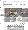
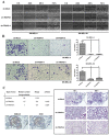
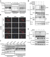
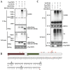
TRAF6 plays a crucial role in the regulation of the innate and adaptive immune responses. Although studies have shown that TRAF6 has oncogenic activity, the role of TRAF6 in melanoma is unclear. Here, we report that TRAF6 is overexpressed in primary as well as metastatic melanoma tumors and melanoma cell lines. Knockdown of TRAF6 with shRNA significantly suppressed malignant phenotypes including cell proliferation, anchorage-independent cell growth and metastasis in vitro and in vivo. Notably, we demonstrated that Basigin (BSG)/CD147, a critical molecule for cancer cell invasion and metastasis, is a novel interacting partner of TRAF6. Furthermore, depletion of TRAF6 by shRNA reduced the recruitment of BSG to the plasma membrane and K63-linked ubiquitination, in turn, which impaired BSG-dependent MMP9 induction. Taken together, our findings indicate that TRAF6 is involved in regulating melanoma invasion and metastasis, suggesting that TRAF6 may be a potential target for therapy or chemo-prevention in melanoma.
Keywords: Basigin; TRAF6; invasion and metastasis; melanoma; ubiquitination.
Conflict of interest statement
No potential conflicts of interest were disclosed.
Figures


Similar articles
-
Epigallocatechin-3-gallate(EGCG) suppresses melanoma cell growth and metastasis by targeting TRAF6 activity.Oncotarget. 2016 Nov 29;7(48):79557-79571. doi: 10.18632/oncotarget.12836. Oncotarget. 2016. PMID: 27791197 Free PMC article.
-
siRNA-induced TRAF6 knockdown promotes the apoptosis and inhibits the invasion of human lung cancer SPC-A1 cells.Oncol Rep. 2016 Apr;35(4):1933-40. doi: 10.3892/or.2016.4602. Epub 2016 Feb 1. Oncol Rep. 2016. PMID: 26847475 Free PMC article.
-
A small interfering CD147-targeting RNA inhibited the proliferation, invasiveness, and metastatic activity of malignant melanoma.Cancer Res. 2006 Dec 1;66(23):11323-30. doi: 10.1158/0008-5472.CAN-06-1536. Cancer Res. 2006. PMID: 17145878
-
CD147/basigin promotes progression of malignant melanoma and other cancers.J Dermatol Sci. 2010 Mar;57(3):149-54. doi: 10.1016/j.jdermsci.2009.12.008. Epub 2010 Jan 8. J Dermatol Sci. 2010. PMID: 20060267 Review.
-
Repressing CD147 is a novel therapeutic strategy for malignant melanoma.Oncotarget. 2017 Apr 11;8(15):25806-25813. doi: 10.18632/oncotarget.15709. Oncotarget. 2017. PMID: 28445958 Free PMC article. Review.
Cited by
-
Matrix Metalloproteinases and Their Inhibitors in Pulmonary Fibrosis: EMMPRIN/CD147 Comes into Play.Int J Mol Sci. 2022 Jun 21;23(13):6894. doi: 10.3390/ijms23136894. Int J Mol Sci. 2022. PMID: 35805895 Free PMC article. Review.
-
The molecular mechanisms of vulpinic acid induced programmed cell death in melanoma.Mol Biol Rep. 2022 Sep;49(9):8273-8280. doi: 10.1007/s11033-022-07619-3. Epub 2022 Aug 12. Mol Biol Rep. 2022. PMID: 35960408
-
An overview of recent molecular dynamics applications as medicinal chemistry tools for the undruggable site challenge.Medchemcomm. 2018 Apr 19;9(6):920-936. doi: 10.1039/c8md00166a. eCollection 2018 Jun 1. Medchemcomm. 2018. PMID: 30108981 Free PMC article. Review.
-
CTHRC1 is associated with BRAF(V600E) mutation and correlates with prognosis, immune cell infiltration, and drug resistance in colon cancer, thyroid cancer, and melanoma.Biomol Biomed. 2024 Dec 11;25(1):42-61. doi: 10.17305/bb.2024.10397. Biomol Biomed. 2024. PMID: 39052013 Free PMC article.
-
Involvement of E3 Ligases and Deubiquitinases in the Control of HIF-α Subunit Abundance.Cells. 2019 Jun 15;8(6):598. doi: 10.3390/cells8060598. Cells. 2019. PMID: 31208103 Free PMC article. Review.
References
-
- Trinh VA. Current management of metastatic melanoma. Am J Health Syst Pharm. 2008;65:S3–8. - PubMed
-
- Simard EP, Ward EM, Siegel R, Jemal A. Cancers with increasing incidence trends in the United States: 1999 through 2008. CA Cancer J Clin. 2012;62:118–128. - PubMed
-
- Cancer Facts & Figures 2014. 2014.
-
- Arch RH, Gedrich RW, Thompson CB. Tumor necrosis factor receptor-associated factors (TRAFs)---a family of adapter proteins that regulates life and death. Genes & Development. 1998;12:2821–2830. - PubMed
Publication types
MeSH terms
Substances
LinkOut - more resources
Full Text Sources
Other Literature Sources
Medical
Molecular Biology Databases
Miscellaneous

