. 2016 Feb 9;7(6):7193-206.
doi: 10.18632/oncotarget.6887.

DANGER is involved in high glucose-induced radioresistance through inhibiting DAPK-mediated anoikis in non-small cell lung cancer

Affiliations

DANGER is involved in high glucose-induced radioresistance through inhibiting DAPK-mediated anoikis in non-small cell lung cancer

TaeWoo Kwon et al. Oncotarget. .

Abstract

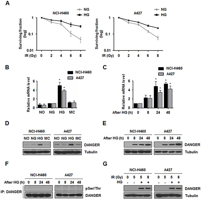
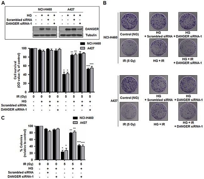
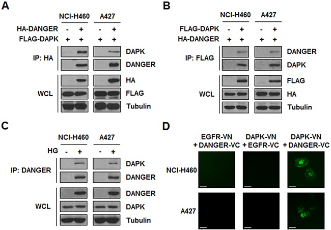
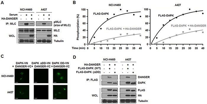
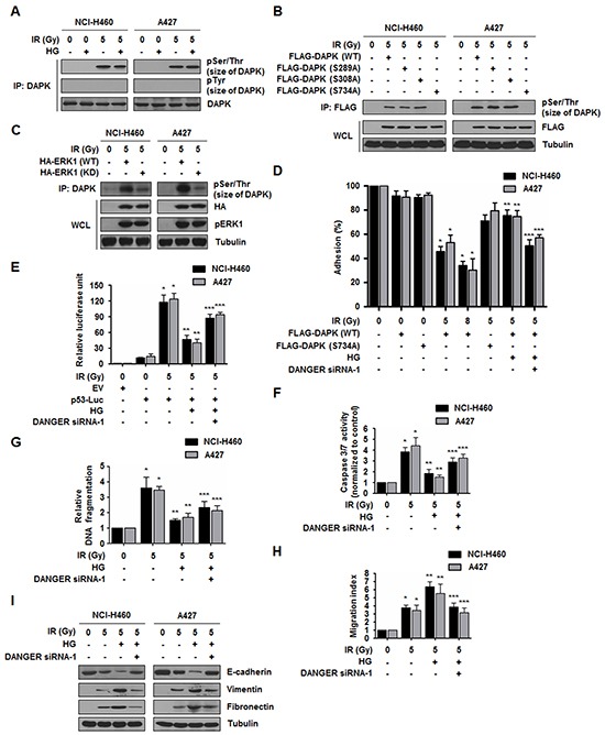
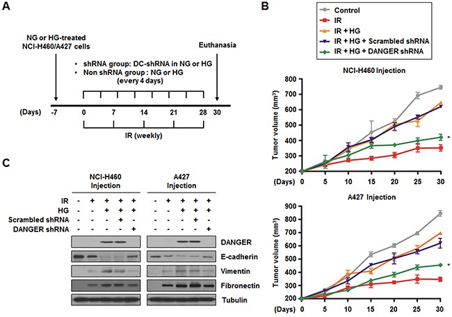
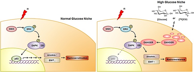
18F-labeled fluorodeoxyglucose (FDG) uptake during FDG positron emission tomography seems to reflect increased radioresistance. However, the exact molecular mechanism underlying high glucose (HG)-induced radioresistance is unclear. In the current study, we showed that ionizing radiation-induced activation of the MEK-ERK-DAPK-p53 signaling axis is required for anoikis (anchorage-dependent apoptosis) of non-small cell lung cancer (NSCLC) cells in normal glucose media. Phosphorylation of DAPK at Ser734 by ERK was essential for p53 transcriptional activity and radiosensitization. In HG media, overexpressed DANGER directly bound to the death domain of DAPK, thus inhibiting the catalytic activity of DAPK. In addition, inhibition of the DAPK-p53 signaling axis by DANGER promoted anoikis-resistance and epithelial-mesenchymal transition (EMT), resulting in radioresistance of HG-treated NSCLC cells. Notably, knockdown of DANGER enhanced anoikis, EMT inhibition, and radiosensitization in a mouse xenograft model of lung cancer. Taken together, our findings offered evidence that overexpression of DANGER and the subsequent inhibitory effect on DAPK kinase activity are critical responses that account for HG-induced radioresistance of NSCLC.

Keywords: DANGER; DAPK; anoikis; high glucose; radioresistance.


Conflict of interest statement

CONFLICTS OF INTEREST

The authors declare no conflict of interest.

Figures

Figure 1
Figure 1. HG induces DANGER overexpression in NSCLC cells
A. Survival curves for NG- or HG-treated NCI-H460 and A427 cells exposed to IR were assessed with a colony forming assay. Data are represented as mean ± SEM (n = 3). B. Expression of ITPRIP in HG-treated NCI-H460 and A427 cells was analyzed by qRT-PCR. After treatment with specific media (NO, no glucose; NG, normal glucose; HG, high glucose; or MC, mannitol control) for 24 h, relative mRNA levels of DANGER were monitored. Data are represented as mean ± SEM (n = 3); *p < 0.05 compared to the NG-treated cells. C. Time-dependent expression of ITPRIP in HG-treated NCI-H460 and A427 cells was analyzed by qRT-PCR. Data are represented as mean ± SEM (n = 3); *p < 0.05 compared with cells after 0 h of HG treatment. D. and E. Expression of DANGER and time-dependent changes in HG-treated NCI-H460 and A427 cells were analyzed by Western blotting. F. HG-induced phosphorylation of DANGER was evaluated with an in vivo kinase assay. After HG treatment for the indicated time, the cells were harvested and cell lysates were subjected to an IP assay with an anti-DANGER antibody followed by Western blotting for pSer/Thr. G. Effect of IR on HG-induced overexpression of DANGER was analyzed by Western blotting.
Figure 2
Figure 2. Down-regulation of DANGER expression reduces HG-induced radioresistance in NSCLC cells
A. Short-term effects of DANGER knockdown on cell growth in both NCI-H460 and A427 cells following IR exposure were assessed with an MTT assay. Data are represented as mean ± SEM (n = 3); *p < 0.05 compared to non-irradiated cells, **p < 0.05 compared to cells treated with irradiation-alone, ***p < 0.05 compared to HG-treated and irradiated cells. Inset: SiRNA knockdown efficiency of DANGER in HG-treated NCI-H460 and A427 cells was analyzed by Western blotting. B. Long-term effects of DANGER knockdown on cell growth in both NCI-H460 and A427 cells following IR exposure were assessed with a colony forming assay. C. Quantitative analysis of the number of NCI-H460 and A427 cell clones after DANGER knockdown with or without HG treatment was performed with Image J. Data are represented as mean ± SEM (n = 3); *p < 0.05 compared to non-irradiated cells, **p < 0.05 compared to cells treated with irradiation-alone, ***p < 0.05 compared to HG-treated and irradiated cells.
Figure 3
Figure 3. HG-induced DANGER physically interacts with DAPK in NSCLC cells
A. and B. Binding of DANGER and DAPK was measured by a reciprocal IP assay. C. HG-induced overexpression of DANGER and subsequent interaction of endogenous DANGER with endogenous DAPK was detected using an IP assay. D. A BiFC assay was performed to evaluate the interaction of DAPK-DANGER in live cells. Cells were transiently transfected with pBiFC-DAPK-VN, pBiFC-DANGER-VC, pBiFC-EGFR-VN, and/or pBiFC-EGFR-VC. Fluorescence indicative of DANGER-DAPK binding was measured in NCI-H460 or A427 cells. Scale bars, 10 μm.
Figure 4
Figure 4. DANGER inhibits the catalytic activity of DAPK in NSCLC cells
A. Effects of DANGER overexpression on DAPK activity were measured with a kinase assay. B. Effects of DANGER on kinetics of MLC phosphorylation by DAPK were measured with a kinetic analysis. The extent of phosphorylation was quantified and expressed as a percentage with 100% representing the maximum phosphorylation of MLC. C. A BiFC assay was performed to determine the interaction of DAPK with DANGER in live cells. NCI-H460 or A427 cells were transiently transfected with pBiFC-DAPK (WT, DD-only, or ΔDD)-VN and/or pBiFC-DANGER-VC. Fluorescence indicative of DANGER-DAPK binding was measured in the cells. Scale bars, 10 μm. D. Involvement of the DAPK DD in binding to DANGER was confirmed with an IP assay.
Figure 5
Figure 5. DANGER reduces DAPK-dependent anoikis in irradiated NSCLC cells
A. HG- and IR-dependent phosphorylation of DAPK was assessed. B. IR-induced DAPK phosphorylation of Ser residues was confirmed using DAPK mutants (S289A, S308A, or S734A). C. Phosphorylation of DAPK by ERK1 was measured using an ERK1-KD (K71R) mutant. pERK1 indicates phosphorylated ERK1. D. Effects of DANGER knockdown on the anti-adhesion effect of DAPK were measured with an adhesion analysis. The cells were irradiated and cell adhesion on fibrinogen was measured. Data are represented as mean ± SEM (n = 3); *p < 0.05 compared to non-irradiated cells transfected with DAPK-WT, **p < 0.05 compared to irradiated cells transfected with DAPK-WT, ***p < 0.05 compared to HG-treated and irradiated cells transfected with DAPK-WT. E. Effects of DANGER knockdown on HG- and IR-induced p53 transcriptional activation were measured with a luciferase assay. Data are represented as mean ± SEM (n = 3); *p < 0.05 compared to non-irradiated cells, **p < 0.05 compared to cells treated with irradiation-alone, ***p < 0.05 compared to HG-treated and irradiated cells. F. Effects of DANGER knockdown on HG- and IR-induced Caspase 3/7 activation in NSCLC cells were measured with a Caspase 3/7 activity assay. Data are represented as mean ± SEM (n = 3); *p < 0.05 compared to non-irradiated cells, **p < 0.05 compared to cells treated with irradiation-alone, ***p < 0.05 compared to HG-treated and irradiated cells. G. Functional involvement of DANGER knockdown in HG- and IR-induced DNA damage responses was measured with a DNA fragmentation assay. Data are represented as mean ± SEM (n = 3); *p < 0.05 compared to non-irradiated cells, **p < 0.05 compared to cells treated with irradiation alone, ***p < 0.05 compared to HG-treated and irradiated cells. H. The inhibitory effects of DANGER knockdown on HG- and IR-induced NSCLC cell migration were measured using a Transwell migration assay. Data are represented as mean ± SEM (n = 3); *p < 0.05 compared to non-irradiated cells, **p < 0.05 compared to cells treated with irradiation-alone, ***p < 0.05 compared to HG-treated and irradiated cells. I. Effects of DANGER knockdown on the protein expression of E-cadherin, Vimentin, and Fibronectin in HG-treated NSCLC cells were analyzed by Western blotting.
Figure 6
Figure 6. Knockdown of DANGER enhances in vivo radiosensitization and decreases in vivo EMT in a xenograft mouse model
A. The experimental protocol for determining whether DANGER knockdown increases in vivo radiosensitization and EMT in a xenograft mouse model (control or DANGER-specific shRNA encapsulated into DOTAP-cholesterol, DC-shRNA).B. The effects of DANGER knockdown by DANGER shRNA-1 on in vivo radiosensitization were measured in a xenograft mouse model. Data are represented as mean ± SEM (n = 3 with three animals/group); *p < 0.05 compared to tumor volume on day-30 in mice treated with radiation, HG, and Scrambled shRNA. C. The in vivo effects of DANGER knockdown by DANGER shRNA-1 on the expression of DANGER and EMT-related proteins were evaluated by Western blot analysis.
Figure 7
Figure 7. A schematic diagram illustrating MEK-ERK-DAPK-p53 signaling in IR-induced anoikis and the radioresistant effect of DANGER in HG-treated NSCLC cells
With NG media, DAPK is phosphorylated on Ser734 by ERK after radiation exposure, consequently leading to the activation of p53 and radiosensitization. IR-induced activation of the MEK-ERK-DAPK-p53 signaling axis is required for anoikis in NSCLC cells. DANGER is overexpressed in NSCLC cells cultured in HG medium, but not in ones maintained under NG conditions. In HG media, overexpressed DANGER directly binds to the DD of DAPK, thus inhibiting the catalytic activity of DAPK. In addition, inhibition of the DAPK-p53 signaling axis by DANGER promotes anoikis-resistance and EMT induction, resulting in radioresistance of HG-treated NSCLC cells. Knockdown of DANGER expression can enhance anoikis, EMT inhibition, and radiosensitization both in vitro and in vivo.

Similar articles

Cited by

References

    1. Koh PK, Faivre-Finn C, Blackhall FH, De Ruysscher D. Targeted agents in non-small cell lung cancer (NSCLC): clinical developments and rationale for the combination with thoracic radiotherapy. Cancer Treat Rev. 2012;38:626–640. - PubMed
    1. Bussink J, van der Kogel AJ, Kaanders JH. Activation of the PI3-K/AKT pathway and implications for radioresistance mechanisms in head and neck cancer. Lancet Oncol. 2008;9:288–296. - PubMed
    1. Nishimura Y, Nakagawa K, Takeda K, Tanaka M, Segawa Y, Tsujino K, Negoro S, Fuwa N, Hida T, Kawahara M, Katakami N, Hirokawa K, Yamamoto N, et al. Phase I/II trial of sequential chemoradiotherapy using a novel hypoxic cell radiosensitizer, doranidazole (PR-350), in patients with locally advanced non-small-cell lung Cancer (WJTOG-0002) Int J Radiat Oncol Biol Phys. 2007;69:786–792. - PubMed
    1. Yang HJ, Youn H, Seong KM, Jin YW, Kim J, Youn B. Phosphorylation of ribosomal protein S3 and antiapoptotic TRAF2 protein mediates radioresistance in non-small cell lung cancer cells. J Biol Chem. 2013;288:2965–2975. - PMC - PubMed
    1. Kim E, Youn H, Kwon T, Son B, Kang J, Yang HJ, Seong KM, Kim W, Youn B. PAK1 tyrosine phosphorylation is required to induce epithelial-mesenchymal transition and radioresistance in lung cancer cells. Cancer Res. 2014;74:5520–5531. - PubMed

Publication types

MeSH terms