. 2016 Mar;22(3):443-55.
doi: 10.1261/rna.054643.115. Epub 2016 Jan 14.

Activating the branch-forming splicing pathway by reengineering the ribozyme component of a natural group II intron

Affiliations

Activating the branch-forming splicing pathway by reengineering the ribozyme component of a natural group II intron

Dario Monachello et al. RNA. 2016 Mar.

Abstract

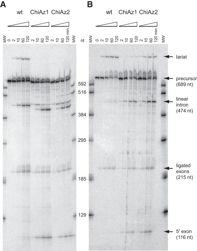
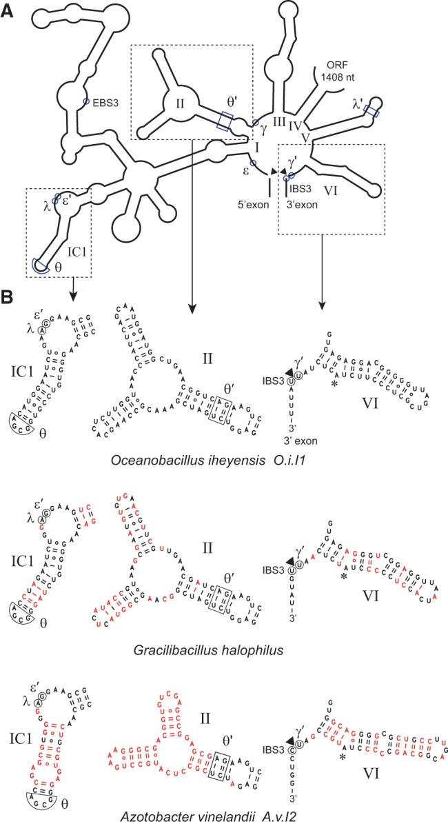
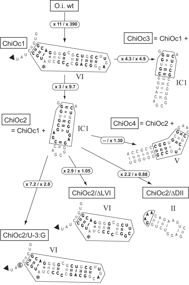
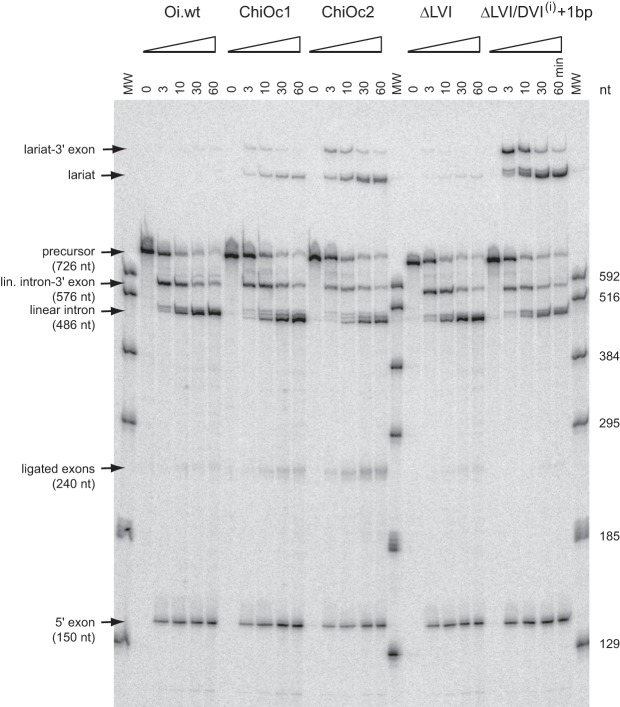
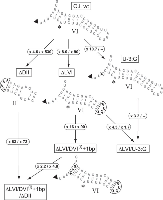
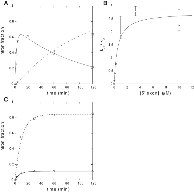
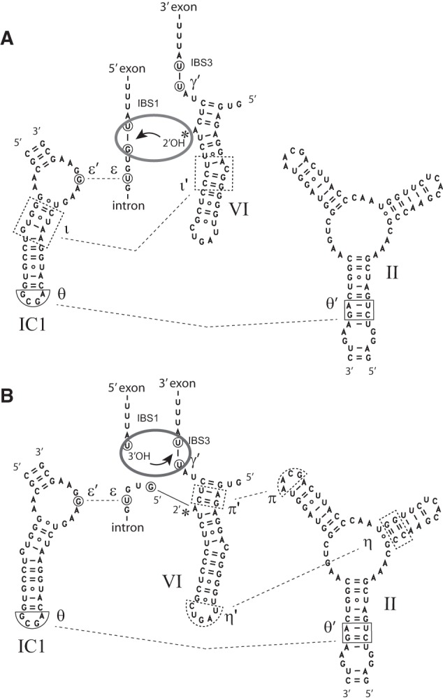
When assayed in vitro, group IIC self-splicing introns, which target bacterial Rho-independent transcription terminators, generally fail to yield branched products during splicing despite their possessing a seemingly normal branchpoint. Starting with intron O.i.I1 from Oceanobacillus iheyensis, whose crystallographically determined structure lacks branchpoint-containing domain VI, we attempted to determine what makes this intron unfit for in vitro branch formation. A major factor was found to be the length of the helix at the base of domain VI: 4 base pairs (bp) are required for efficient branching, even though a majority of group IIC introns have a 3-bp helix. Equally important for lariat formation is the removal of interactions between ribozyme domains II and VI, which are specific to the second step of splicing. Conversely, mismatching of domain VI and its proposed first-step receptor in subdomain IC1 was found to be detrimental; these data suggest that the intron-encoded protein may promote branch formation partly by modulating the equilibrium between conformations specific to the first and second steps of splicing. As a practical application, we show that by making just two changes to the O.i.I1 ribozyme, it is possible to generate sufficient amounts of lariat intron for the latter to be purified and used in kinetic assays in which folding and reaction are uncoupled.

Keywords: group II intron; lariat intron; linear intron; self-splicing.


Figures

FIGURE 1.
FIGURE 1.
Time course of self-splicing at 55°C of intron A.v.I2 and A.v.I2-based chimeras ChiAz1 and ChiAz2. (A) Magnesium-containing buffer; (B) Manganese-containing buffer (see Materials and Methods for full experimental conditions). Bands corresponding to linear products were identified by reference to transcripts of known length (lanes MW).
FIGURE 2.
FIGURE 2.
Phylogenetic tree of bacterial class C introns based on alignment of intron-encoded protein sequences (see Materials and Methods). Whenever possible, intron names are abbreviated as in previous works (Candales et al. 2012; Toro and Martínez-Abarca 2013). Arrows point to introns whose self-splicing reaction was examined in vitro (Granlund et al. 2001; Toor et al. 2006, , ; this work), circled +1 signs indicate clades whose intron members have acquired an additional (fourth) base pair in their DVI basal helix (according to secondary structure models in the Database for bacterial group II introns of Candales et al. [2012] and F Michel [data not shown]).
FIGURE 3.
FIGURE 3.
(A) Organization of secondary structure domains I to VI of intron O.i.I1 (from Oceanobacillus iheyensis). (B) Detailed secondary structure of domains IC1, II, and VI in introns O.i.I1, A.v.I2, and Gracilibacillus halophilus. Nucleotides that differ from their counterparts in O.i.I1 are in red. The asterisk marks the predicted branchpoint and an arrowhead points to the expected intron–3′ exon junction. Greek letters, EBS3, and IBS3 indicate nucleotides that participate in tertiary interactions (Costa et al. 2000).
FIGURE 4.
FIGURE 4.
Chimeras based on the Oceanobacillus intron. Changes introduced with respect to the O.i.I1 sequence are in bold type. Arrows indicate the genealogy of constructs, e.g., chimera ChiOc2 was derived from ChiOc1 through replacement of O.i.I1 IC1 by its counterpart in A.v.I2. Circled numbers are increases in the ratio of initial rates of branching over hydrolysis (Table 1) that result from the molecular alterations shown in the figure (values for magnesium- and manganese-containing buffers are to the left and right side of the slash, respectively).
FIGURE 5.
FIGURE 5.
Time course of self-splicing of O.i.I1-based chimeras and mutants (see Figs. 4, 6) in manganese-containing buffer (see Materials and Methods). Bands corresponding to linear products were identified by reference to transcripts of known length (lanes MW). Multiple bands visible at short reaction times just above the lariat intron of construct ΔLVI/DVI(i) + 1 bp were assumed to have been generated by the same process that results in multiple linear intron forms: Based on the migration of the latter, those bands were tentatively attributed to the use of surrogate 3′ splice sites after positions +7 and +14 of the 3′ exon.
FIGURE 6.
FIGURE 6.
Modifications made to the Oceanobacillus intron. Changes are boxed; circled numbers as in Figure 4.
FIGURE 7.
FIGURE 7.
Ratios of initial rate of branching over hydrolysis in manganese- (Rbr/hy,Mn) versus magnesium-containing (Rbr/hy,Mg) buffers. Constructs are depicted in Figures 4 and 6. Bars indicate standard errors (see Materials and Methods). The distribution of data points was tentatively fitted to a log-log linear equation (line).
FIGURE 8.
FIGURE 8.
Analysis of reaction kinetics. (A) Time course of debranching of purified ΔLVI/DVI(i) + 1 bp lariat molecules by 10 μM oligonucleotide Oceano 14-6 (see Materials and Methods). Circles and full curve, E5–intron linear form; squares, intron linear form. Reaction conditions: 45°C, 2 M NaCl, 40 mM Na-HEPES, pH 7.0 (37°C), 10 mM MnCl2, 0.01% SDS, 20 nM lariat. Debranched and linear fractions were fitted with Kaleidagraph 3.6 to Equations 1 and 2 in Materials and Methods, respectively, with 1/τ1 = 0.410 ± 0.046 min−1, 1/τ2 = 0.0098 ± 0.0011 min−1, and kdb = 0.285 ± 0.024 min−1 (Pearson's R was 0.9947 and 0.9967, respectively). (B) Debranching equilibrium of the ΔLVI/DVI(i) + 1 bp lariat as a function of the concentration of oligonucleotide Oceano 14-6. Reaction conditions: 45°C, 2 M NH4Cl, 50 mM Tris-HCl, pH 7.5 (37°C), 10 mM MnCl2, 0.01% SDS, 10 nM lariat. Bars indicate standard errors. See Materials and Methods for calculation of kdb and kbr. Data were fitted with Kaleidagraph 3.6 to (kdb,max/kbr)/(1 + Kd/[E5]), with kdb,max/kbr = 2.7 ± 0.03 and Kd = 0.56 ± 0.22 μM. (C) Reaction kinetics of precursor transcript O.i.I1 ΔDII in manganese-containing buffer. Data were fitted with Kaleidagraph 3.6 to (v0/k) [1−exp(−kt)], where k is the rate constant and v0 the initial rate of reaction. Circles and full curve, fraction of branched intron products (k = 0.133 ± 0.011 min−1, v0 = 0.015 ± 0.001 min−1); squares and dashed curve, fraction of linear intron products (k = 0.097 ± 0.007 min−1, v0 = 0.082 ± 0.006 min−1).
FIGURE 9.
FIGURE 9.
Postulated displacement of domain VI between the two splicing steps. (A) First-step conformation. (B) Second-step conformation. The θ–θ′ and ε–ε′ interactions (Costa et al. 1997a; Toor et al. 2008) are presumed to persist throughout the splicing process, whereas ι–ι′, η–η′, and π–π′ are step-specific interactions, whose location in the Oceanobacillus intron is tentatively inferred from studies on other group II introns (Li et al. 2011b; Robart et al. 2014). A gray ellipse symbolizes the catalytic center of the group II ribozyme and the arrow indicates nucleophilic attack of a phosphodiester bond. Movement of domain VI between its two preferred locations is assumed to occur irrespective of the presence of the 5′ exon (see text).

Similar articles

Cited by

References

    1. Adamidi C, Fedorova O, Pyle AM. 2003. A group II intron inserted into a bacterial heat-shock operon shows autocatalytic activity and unusual thermostability. Biochemistry 42: 3409–3418. - PubMed
    1. Belfort M, Roberts RJ. 1997. Homing endonucleases: keeping the house in order. Nucleic Acids Res 25: 3379–3388. - PMC - PubMed
    1. Bell-Pedersen D, Quirk S, Clyman J, Belfort M. 1990. Intron mobility in phage T4 is dependent upon a distinctive class of endonucleases and independent of DNA sequences encoding the intron core: mechanistic and evolutionary implications. Nucleic Acids Res 18: 3763–3770. - PMC - PubMed
    1. Candales MA, Duong A, Hood KS, Li T, Neufeld RAE, Sun R, McNeil BA, Wu L, Jarding AM, Zimmerly S. 2012. Database for bacterial group II introns. Nucleic Acids Res 40: D187–D190. - PMC - PubMed
    1. Chanfreau G, Jacquier A. 1996. An RNA conformational change between the two chemical steps of group II self-splicing. EMBO J 15: 3466–3476. - PMC - PubMed

Publication types

LinkOut - more resources