

Blue laser imaging endoscopy system for the early detection and characterization of colorectal lesions: a guide for the endoscopist
- PMID: 26770267
- PMCID: PMC4699272
- DOI: 10.1177/1756283X15603614
Blue laser imaging endoscopy system for the early detection and characterization of colorectal lesions: a guide for the endoscopist
Abstract
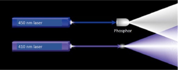
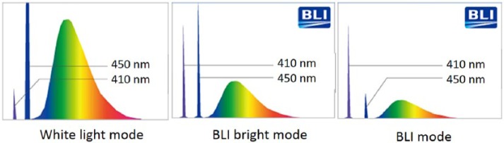
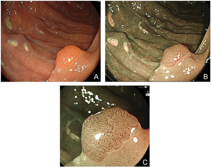
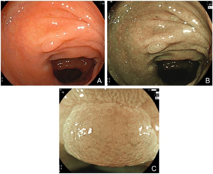
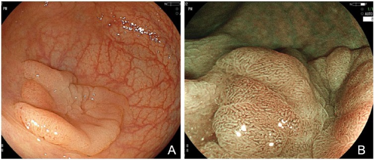
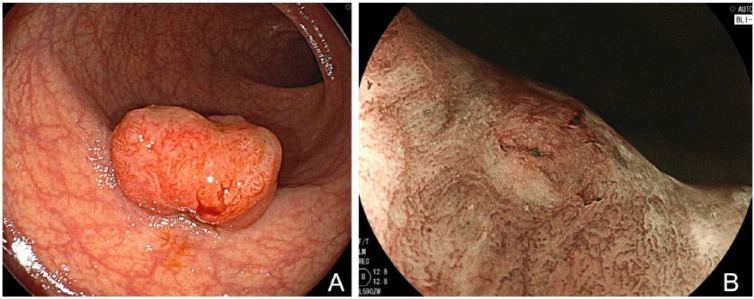
Blue laser imaging is a new system for image-enhanced endoscopy using laser light. Blue laser imaging utilizes two monochromatic lasers (410 and 450 nm) instead of xenon light. A 410 nm laser visualizes vascular microarchitecture, similar to narrow band imaging, and a 450 nm laser provides white light by excitation. According to three recently published reports, the diagnostic ability of polyp characterization using blue laser imaging compares favorably with narrow band imaging. No published data are available to date regarding polyp detection with blue laser imaging. However, blue laser imaging has the possibility to increase the detection of colorectal polyps by depicting brighter and clearer endoscopic images, even at a distant view, compared with first-generation image-enhanced endoscopy. A clinical trial to compare the detection between blue laser imaging and xenon light is warranted.
Keywords: blue laser imaging; colon polyp; colorectal cancer; colorectal neoplasm; image enhanced endoscopy; magnification colonoscopy.
Conflict of interest statement
Figures
References
-
- Adler A., Aschenbeck J., Yenerim T., Mayr M., Aminalai A., Drossel R., et al. (2009) Narrow-band versus white-light high definition television endoscopic imaging for screening colonoscopy: a prospective randomized trial. Gastroenterology 136: 410–416 e411; quiz 715. - PubMed
-
- Aminalai A., Rosch T., Aschenbeck J., Mayr M., Drossel R., Schroder A., et al. (2010) Live image processing does not increase adenoma detection rate during colonoscopy: a randomized comparison between FICE and conventional imaging (Berlin Colonoscopy Project 5, BECOP-5). Am J Gastroenterol 105: 2383–2388. - PubMed
-
- Axelrad A., Fleischer D., Geller A., Nguyen C., Lewis J., Al-Kawas F., et al. (1996) High-resolution chromoendoscopy for the diagnosis of diminutive colon polyps: implications for colon cancer screening. Gastroenterology 110: 1253–1258. - PubMed
-
- Hoffman A., Kagel C., Goetz M., Tresch A., Mudter J., Biesterfeld S., et al. (2010a) Recognition and characterization of small colonic neoplasia with high-definition colonoscopy Using i-SCAN is as precise as chromoendoscopy. Dig Liver Dis 42: 45–50. - PubMed
-
- Hoffman A., Sar F., Goetz M., Tresch A., Mudter J., Biesterfeld S., et al. (2010b) High definition colonoscopy combined with i-SCAN is superior in the detection of colorectal neoplasias compared with standard video colonoscopy: a prospective randomized controlled trial. Endoscopy 42: 827–833. - PubMed
Publication types
LinkOut - more resources
Full Text Sources
Medical
