

Trauma of the midface
- PMID: 26770280
- PMCID: PMC4702055
- DOI: 10.3205/cto000121
Trauma of the midface
Abstract
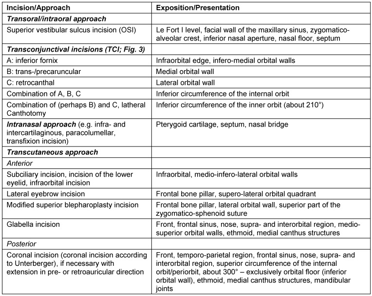
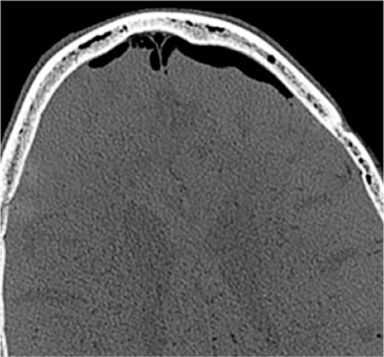
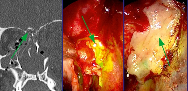
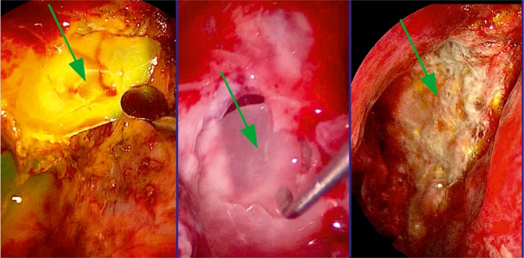
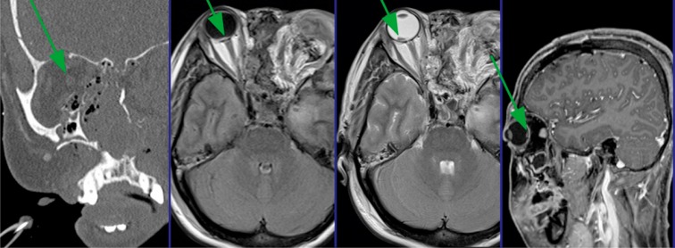
Fractures of the midface pose a serious medical problem as for their complexity, frequency and their socio-economic impact. Interdisciplinary approaches and up-to-date diagnostic and surgical techniques provide favorable results in the majority of cases though. Traffic accidents are the leading cause and male adults in their thirties are affected most often. Treatment algorithms for nasal bone fractures, maxillary and zygomatic fractures are widely agreed upon whereas trauma to the frontal sinus and the orbital apex are matter of current debate. Advances in endoscopic surgery and limitations of evidence based gain of knowledge are matters that are focused on in the corresponding chapter. As for the fractures of the frontal sinus a strong tendency towards minimized approaches can be seen. Obliteration and cranialization seem to decrease in numbers. Some critical remarks in terms of high dose methylprednisolone therapy for traumatic optic nerve injury seem to be appropriate. Intraoperative cone beam radiographs and preshaped titanium mesh implants for orbital reconstruction are new techniques and essential aspects in midface traumatology. Fractures of the anterior skull base with cerebrospinal fluid leaks show very promising results in endonasal endoscopic repair.
Keywords: ESS; midface fractures; orbit; rhinology; trauma.
Figures
References
-
- Maier H, Tisch M, Lorenz KJ, Danz B, Schramm A. Penetrierende Gesichts- und Halsverletzungen. Diagnostik und Therapie. [Penetrating injuries in the face and neck region. Diagnosis and treatment]. HNO. 2011 Aug;59(8):765–782. doi: 10.1007/s00106-011-2349-1. (Ger). Available from: http://dx.doi.org/10.1007/s00106-011-2349-1. - DOI - PubMed
-
- Frodel JL., Jr Avoiding and correcting complications in perinasal trauma. Facial Plast Surg. 2012 Jun;28(3):323–332. doi: 10.1055/s-0032-1312697. Available from: http://dx.doi.org/10.1055/s-0032-1312697. - DOI - PubMed
-
- Alvi A, Doherty T, Lewen G. Facial fractures and concomitant injuries in trauma patients. Laryngoscope. 2003 Jan;113(1):102–106. doi: 10.1097/00005537-200301000-00019. Available from: http://dx.doi.org/10.1097/00005537-200301000-00019. - DOI - PubMed
-
- Buchanan EP, Hopper RA, Suver DW, Hayes AG, Gruss JS, Birgfeld CB. Zygomaticomaxillary complex fractures and their association with naso-orbito-ethmoid fractures: a 5-year review. Plast Reconstr Surg. 2012 Dec;130(6):1296–1304. doi: 10.1097/PRS.0b013e31826d1643. Available from: http://dx.doi.org/10.1097/PRS.0b013e31826d1643. - DOI - PubMed
-
- Walters JT, Bisson JI, Shepherd JP. Predicting post-traumatic stress disorder: validation of the Trauma Screening Questionnaire in victims of assault. Psychol Med. 2007 Jan;37(1):143–150. doi: 10.1017/S0033291706008658. Available from: http://dx.doi.org/10.1017/S0033291706008658. - DOI - PubMed
Publication types
LinkOut - more resources
Full Text Sources
Medical
Research Materials
Miscellaneous
