Review
. 2015 Dec 22:14:Doc08.
doi: 10.3205/cto000123. eCollection 2015.

Comprehensive review on endonasal endoscopic sinus surgery

Affiliations
Review

Comprehensive review on endonasal endoscopic sinus surgery

Rainer K Weber et al. GMS Curr Top Otorhinolaryngol Head Neck Surg. .

Abstract

Endonasal endoscopic sinus surgery is the standard procedure for surgery of most paranasal sinus diseases. Appropriate frame conditions provided, the respective procedures are safe and successful. These prerequisites encompass appropriate technical equipment, anatomical oriented surgical technique, proper patient selection, and individually adapted extent of surgery. The range of endonasal sinus operations has dramatically increased during the last 20 years and reaches from partial uncinectomy to pansinus surgery with extended surgery of the frontal (Draf type III), maxillary (grade 3-4, medial maxillectomy, prelacrimal approach) and sphenoid sinus. In addition there are operations outside and beyond the paranasal sinuses. The development of surgical technique is still constantly evolving. This article gives a comprehensive review on the most recent state of the art in endoscopic sinus surgery according to the literature with the following aspects: principles and fundamentals, surgical techniques, indications, outcome, postoperative care, nasal packing and stents, technical equipment.

Keywords: FESS; endoscopic sinus surgery; nasal packing; outcome after sinus surgery; postoperative care after sinus surgery.


Figures
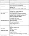
Table 1
Table 1. CT checklist before sinus surgery
Table 2
Table 2. Staging classification of chronic rhinosinusitis according to Kennedy (based on CT scans)
Table 3
Table 3. Classification of frontal sinus operations (FSO, modified classification according to Draf)
Table 4
Table 4. Advantages and disadvantages of endonasal endoscopic procedures for treatment of sinonasal tumors [9]
Table 5
Table 5. Recommendations for the use of navigation systems in the context of endonasal endoscopic sinus surgery according to the current literature [417]
Table 6
Table 6. Recommendations on the basic standard postoperative care after endonasal endoscopic sinus surgery
Figure 1
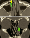
Figure 1. Extended maximal middle meatal antrostomy grade 4 (postlacrimal approach, [293]): Resection of the bone (green) medially, dorsally, and laterally of the nasolacrimal duct in order to mobilize it and to improve the insight into the maxillary sinus. Resection of the bone in case of prelacrimal access (yellow). a) axial CT scan, b) coronal CT scan.
Figure 2
Figure 2. Prelacrimal approach: endoscopic view into the left maxillary sinus. The suction device points at the posterior wall of the maxillary sinus. 1 = nasolacrimal duct, 2 = anterior wall of the maxillary sinus, 3 = alveolar recess.
Figure 3
Figure 3. Missed ostium sequence. a) Typical secretion drop directly behind obvious remnants of the uncinate process in MOS of the right side after previous surgery. b) In the coronal CT scan a larger opening of the maxillary sinus is seen in the posterior part of the middle meatus (*). c) In the area of the natural ostium, however, remnants of the uncinate process and soft tissue are revealed (mucosal swelling, scars (=1) with obstruction of the natural ostium in contrast to free drainage on the left side (=2)).
Figure 4
Figure 4. Sagittal CT demonstrating the surgical strategy to open the posterior ethmoid. After opening the basal lamella of the middle turbinate (1) directly above the horizontal part (2), the superior meatus (3) is reached. (4) = ethmoid bulla.
Figure 5
Figure 5. Transpterygoid approach to the left sphenoid sinus with view into the lateral recess (1), the maxillary nerve that is partly not covered by bone (2), and a part of the middle cranial fossa (3).
Figure 6
Figure 6. Frontal sinus drainage type I according to Draf = complete resection of the uncinate process and resection of parts of the medial lamella of the agger nasi cell and the anterior wall of the ethmoid bulla if needed [242, 246, 248, 359]. A different postoperative situation results depending on the individual anatomy: on the right isolated agger nasi cell, on the left side additional posterior frontoethmoidal cell (frontal bulla), intersinus septal cell; 1 = agger nasi cell, 2 = posterior frontoethmoidal cell (frontal bulla), 3 = interfrontal sinus septal cell, 4 = ethmoid bulla; a) coronal CT scan, b) axial CT scan, c) sagittal CT scan.
Figure 7
Figure 7. Extent of the resection of endonasal endoscopic frontal sinus drainage type IIa, IIb, III according to Draf.
Type IIa = resection of all anterior ethmoid cells obstructing the frontal sinus drainage pathway. Type IIb = type IIa + resection of the ipsilateral floor of the frontal sinus + the ipsilateral middle turbinate in front of the level of the posterior wall of the frontal sinus. Advanced type IIb = type IIb + resection of the frontal sinus septum (blue). Modified type III = type IIb + resection of the nasal septum (green) (+ resection of the contralateral medial floor of the frontal sinus (red) + resection of the complete contralateral floor of the frontal sinus and the contralateral middle turbinate in front of the level of the posterior wall of the frontal sinus if needed (yellow) (if present) + resection of the frontal sinus septum (blue) if needed). Type III = bilateral type IIb + resection of the adjacent nasal septum + resection of the frontal sinus septum.
Figure 8
Figure 8. Condition after frontal sinus drainage type III with maximal opening, smooth transition and coverage of the bare bone with free mucosal transplants (=1)
Figure 9
Figure 9. Exostoses of the frontal sinus, endoscopic view (a) and CT scan (b)
Figure 10
Figure 10. Female patient with condition after pneumococci meningitis and bony defect of the posterior wall of the sphenoid sinus (CT scan) as well as liquor passage into the sphenoid sinus (MRI). a) axial CT scan. b) MRI (CISS sequence).
Figure 11
Figure 11. Female patient with pneumococci meningitis and acute sphenoid sinusitis and intact skull base in thin layer CT scan. Intraoperatively, a bony defect measuring 1–2 mm is obvious at the posterior wall of the sphenoid sinus/anterior wall of the pituitary.
Figure 12
Figure 12. Chronic maxillary sinusitis of the right side. The causative preapical abscess was not identified neither by the dentist nor by the radiologist (CT scan). The papillomatous thickened mucosa can and should be preserved (intraoperative endoscopy, 45° optics).
Figure 13
Figure 13. Silent sinus syndrome of the right maxillary sinus with a small maxillary sinus, lowered floor of the orbit and major retraction of the uncinate process that is directly close to the lamina papyracea
Figure 14
Figure 14. Inverted papilloma of the left maxillary sinus (Krouse stage III). a) Complete opacification of the maxillary sinus by exophytic tumor mass with typical hyperostosis at the small-sized origin at the base of the maxillary sinus (white arrow). b) After resection of the exophytic tumor mass the region of origin is exposed via a prelacrimal approach. Removal of mucosa with 1–1.5 cm safety margins and removal of the bone lying under the tumorattachment. Additional coagulation of an artery of the underlying mucosa of the hard palate. 1 = anterior wall of the maxillary sinus, 2 = region of the removed tumor-affected bone with coagulation after arterial bleeding, 3 = medialized inferior turbinate with nasolacrimal duct. c) Condition after repositioning and fixation of the inferior turbinate. The maxillary sinus can be well accessed via medial maxillectomy for endoscopic control.
Figure 15
Figure 15. Occlusion of the nose for optimized wound healing

References

    1. Draf W. Die chirurgische Behandlung entzündlicher Erkrankungen der Nasennebenhöhlen. Indikation, Operationsverfahren, Gefahren, Fehler und Komplikationen, Revisionschirurgie. [Surgical treatment of the inflammatory diseases of the paranasal sinuses. Indication, surgical technique, risks, mismanagement and complications, revision surgery]. Arch Otorhinolaryngol. 1982;235(1):133–305. (Ger). - PubMed
    1. Hosemann W. Die endonasale Chirurgie der Nasennebenhöhlen – Konzepte, Techniken, Ergebnisse, Komplikationen, Revisionseingriffe. [Endonasal surgery of the paranasal sinuses - concepts, techniques, results, complications and revision interventions]. Eur Arch Otorhinolaryngol Suppl. 1996;1:155–269. (Ger). - PubMed
    1. Stuck BA, Bachert C, Federspil P, Hosemann W, Klimek L, Mösges R, Pfaar O, Rudack C, Sitter H, Wagenmann M, Weber R, Hörmann K German Society of Otorhinolaryngology, Head and Neck Surgery. Leitlinie "Rhinosinusitis" - Langfassung: S2-Leitlinie der Deutschen Gesellschaft für Hals-Nasen-Ohren-Heilkunde, Kopf- und Hals-Chirurgie. [Rhinosinusitis guidelines - unabridged version: S2 guidelines from the German Society of Otorhinolaryngology, Head and Neck Surgery]. HNO. 2012 Feb;60(2):141–162. doi: 10.1007/s00106-011-2396-7. (Ger). Available from: http://dx.doi.org/10.1007/s00106-011-2396-7. - DOI - DOI - PubMed
    1. Hosemann W, Schroeder HW. Comprehensive review on rhino-neurosurgery. GMS Curr Top Otorhinolaryngol Head Neck Surg. 2015;14:Doc01. doi: 10.3205/cto000116. Available from: http://dx.doi.org/10.3205/cto000116. - DOI - DOI - PMC - PubMed
    1. Kühnel TS, Reichert TE. Trauma of the midface. GMS Curr Top Otorhinolaryngol Head Neck Surg. 2015;14:Doc06. doi: 10.3205/cto000121. Available from: http://dx.doi.org/10.3205/cto000121. - DOI - DOI - PMC - PubMed