

Expressions of lysophosphatidic acid receptors in the development of human ovarian carcinoma
- PMID: 26770382
- PMCID: PMC4694282
Expressions of lysophosphatidic acid receptors in the development of human ovarian carcinoma
Abstract
Aim: To investigate the associations between the expressions of three lysophosphatidic acid (LPA) receptors (LPA1-3) and the development of ovarian carcinoma (OC).
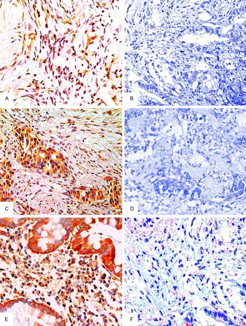
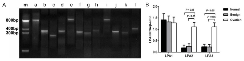
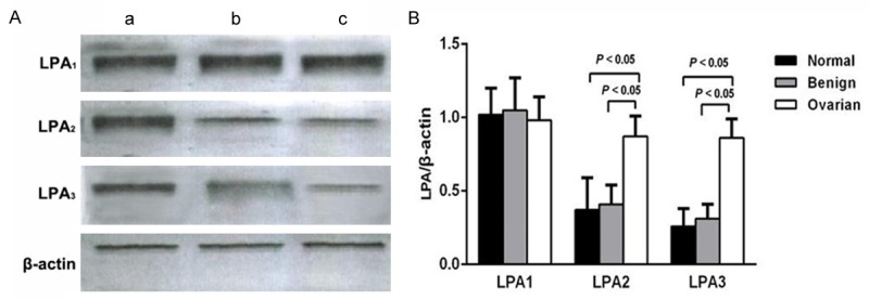
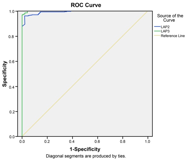
Method: Ovarian tissue specimens, including normal ovarian epithelium tissues, benign ovarian tumor tissues and OC tissues were collected from patients who underwent surgical resections between March 2012 and December 2014. Immunohistochemical staining was used to detect LPA receptor expressions in ovarian tissues. Reverse transcription-polymerase chain reaction and Western blotting were used to detect mRNA and protein expression of LPA receptors, respectively. Association analysis between LPA receptors protein expression and clinical pathological characteristics was conducted. The value of LPA2 and LPA3 in discriminating OC was confirmed by receiver-operator characteristic (ROC) curves analysis.
Results: The positive expression rates of LPA2 and LPA3 in OC group was obviously higher than normal control and benign groups. The LPA2 and LPA3 mRNA and protein levels in OC group were higher than in normal control and benign groups. LPA2 and LPA3 mRNA expression levels were positively correlated with LPA2 and LPA3 protein expression in OC group. ROC curve analysis revealed that LPA2 yield a specificity of 96.3% and a sensitivity of 97.9%, and LPA3 yield a specificity of 98.5% and a sensitivity of 97.9% for the detection of OC.
Conclusion: LPA2 and LPA3 were highly expressed in OC tissues, which may be involved in the development of OC. Further, LPA2 and LPA3 had higher sensitivity and specificity in distinguishing the OC from benign ovarian tumors, which could be potential diagnostic indictors in OC.
Keywords: Ovarian carcinoma; ROC curve analysis; immunohistochemical staining; lysophosphatidic acid receptors; pathological grades; pathological stages; pathological types; reverse transcription-polymerase chain reaction.
Figures
Similar articles
-
Lysophosphatidic acid receptors determine tumorigenicity and aggressiveness of ovarian cancer cells.J Natl Cancer Inst. 2008 Nov 19;100(22):1630-42. doi: 10.1093/jnci/djn378. Epub 2008 Nov 11. J Natl Cancer Inst. 2008. PMID: 19001604 Free PMC article.
-
Aberrant expression of lysophosphatidic acid (LPA) receptors in human colorectal cancer.Lab Invest. 2004 Oct;84(10):1352-62. doi: 10.1038/labinvest.3700146. Lab Invest. 2004. PMID: 15220934
-
Differential targeting of lysophosphatidic acid LPA1, LPA2, and LPA3 receptor signalling by tricyclic and tetracyclic antidepressants.Eur J Pharmacol. 2023 Nov 15;959:176064. doi: 10.1016/j.ejphar.2023.176064. Epub 2023 Sep 25. Eur J Pharmacol. 2023. PMID: 37758013
-
The LPA receptors.Prostaglandins Other Lipid Mediat. 2001 Apr;64(1-4):21-32. doi: 10.1016/s0090-6980(01)00105-8. Prostaglandins Other Lipid Mediat. 2001. PMID: 11324705 Review.
-
Two pathways for lysophosphatidic acid production.Biochim Biophys Acta. 2008 Sep;1781(9):513-8. doi: 10.1016/j.bbalip.2008.06.005. Epub 2008 Jun 24. Biochim Biophys Acta. 2008. PMID: 18621144 Review.
Cited by
-
Dual role of autotaxin as novel biomarker and therapeutic target in pancreatic neuroendocrine neoplasms.Cancer Sci. 2023 Dec;114(12):4571-4582. doi: 10.1111/cas.15980. Epub 2023 Sep 28. Cancer Sci. 2023. PMID: 37770812 Free PMC article.
-
Role of lysophosphatidic acid and its receptors in health and disease: novel therapeutic strategies.Signal Transduct Target Ther. 2021 Feb 1;6(1):45. doi: 10.1038/s41392-020-00367-5. Signal Transduct Target Ther. 2021. PMID: 33526777 Free PMC article. Review.
References
-
- Menon U. Ovarian cancer screening has no effect on disease-specific mortality. Evid Based Med. 2012;17:47–48. - PubMed
-
- Buys SS, Partridge E, Black A, Johnson CC, Lamerato L, Isaacs C, Reding DJ, Greenlee RT, Yokochi LA, Kessel B, Crawford ED, Church TR, Andriole GL, Weissfeld JL, Fouad MN, Chia D, O’Brien B, Ragard LR, Clapp JD, Rathmell JM, Riley TL, Hartge P, Pinsky PF, Zhu CS, Izmirlian G, Kramer BS, Miller AB, Xu JL, Prorok PC, Gohagan JK, Berg CD, Team PP. Effect of screening on ovarian cancer mortality: the Prostate, Lung, Colorectal and Ovarian (PLCO) Cancer Screening Randomized Controlled Trial. JAMA. 2011;305:2295–2303. - PubMed
-
- Orfanelli T, Jeong JM, Doulaveris G, Holcomb K, Witkin SS. Involvement of autophagy in cervical, endometrial and ovarian cancer. Int J Cancer. 2014;135:519–528. - PubMed
-
- Bordon Y. Immunotherapy: Leukadherins get a grip on inflammation. Nat Rev Immunol. 2011;11:638. - PubMed
LinkOut - more resources
Full Text Sources
Miscellaneous