

Application value of computer assisted surgery system in precision surgeries for pediatric complex liver tumors
- PMID: 26770445
- PMCID: PMC4694345
Application value of computer assisted surgery system in precision surgeries for pediatric complex liver tumors
Abstract
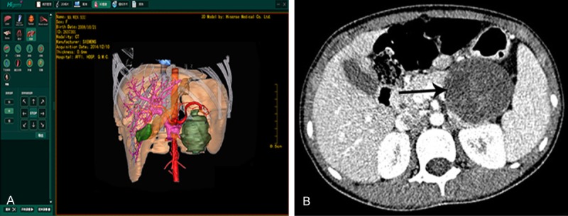
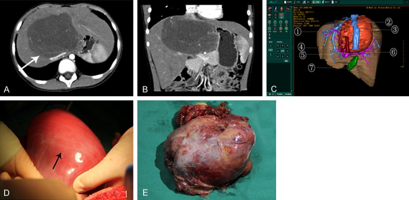
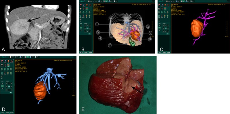
We discussed the diagnostic and treatment value and clinical significance of computer assisted surgery system (Higemi) in precision surgeries for pediatric complex liver tumors. A total of 21 pediatric cases receiving hepatectomy for tumors in the portal vein and giant liver tumors from June 2012 to January 2015 were analyzed. Higemi was used for 3-dimensional (3D) reconstruction of thin-slice CT images and surgical planning. Tumors were precisely located and blood vessel neighborhood was determined so as to evaluate surgical feasibility. In addition, pathological classification, surgical time, intraoperative blood loss, transfusion rate and complications were predicted. After 3D reconstruction using Higemi, the neighboring relationship of tumors with blood vessels and the running direction of the blood vessels were clearly visualized. Of 21 cases, 10 cases had tumors located in the left lobe, 5 cases in the right lobe, 3 cases showing involvement of right trilobes, and 3 cases in the middle lobe. Lobes exceeding one third of the total liver volume were resected in 18 cases. Postoperative pathological examination indicated 10 cases of hepatoblastoma, 3 cases of hepatocellular carcinoma, 3 cases of hamartoma, 3 cases of infantile hemangioendothelioma, 1 case of teratoma and 1 case of undifferentiated malignant mesenchymoma. The surgical time was 90-240 min with an average of 130 min; the medium intraoperative blood loss was 60 ml and the minimum blood loss was 3 ml; the transfusion rate was 42.9% (9/21). Surgeries were successful in 20 cases, who were discharged after recovery. However, one case had giant liver tumor combined with severe obstructive jaundice and hepatic insufficiency and died of postoperative liver failure and DIC. 3D reconstruction of CT data using Higemi can clearly visualize the running direction of blood vessels and the neighboring relationship with tumors. Higemi can improve the precision and safety of complex hepatectomy.
Keywords: 3D; Surgery; computer assisted; imaging; liver resection.
Figures
Similar articles
-
Clinical application of a three-dimensional imaging technique in infants and young children with complex liver tumors.Pediatr Surg Int. 2016 Apr;32(4):387-95. doi: 10.1007/s00383-016-3864-7. Epub 2016 Jan 25. Pediatr Surg Int. 2016. PMID: 26809670
-
[Application of 3D visualization, 3D printing and 3D laparoscopy in the diagnosis and surgical treatment of hepatic tumors].Nan Fang Yi Ke Da Xue Xue Bao. 2015 May;35(5):639-45. Nan Fang Yi Ke Da Xue Xue Bao. 2015. PMID: 26018255 Chinese.
-
[Application of liver three-dimensional visualization technologies in the treatment planning of hepatic malignant tumor].Zhonghua Wai Ke Za Zhi. 2017 Dec 1;55(12):916-922. doi: 10.3760/cma.j.issn.0529-5815.2017.12.008. Zhonghua Wai Ke Za Zhi. 2017. PMID: 29224266 Chinese.
-
A Left-Sided Approach for Resection of Hepatic Caudate Lobe Hemangioma: Two Case Reports and a Literature Review.Int Surg. 2015 Jun;100(6):1054-9. doi: 10.9738/INTSURG-D-14-00317.1. Int Surg. 2015. PMID: 26414827 Free PMC article. Review.
-
Hepatic surgery for metastases from neuroendocrine tumors.Surg Oncol Clin N Am. 2003 Jan;12(1):231-42. doi: 10.1016/s1055-3207(02)00076-5. Surg Oncol Clin N Am. 2003. PMID: 12735141 Review.
Cited by
-
Effective preoperative abdominal incision planning on a patient with a history of repeated abdominal operations using a three-dimensional reconstruction technique: a case report.J Int Med Res. 2019 Mar;47(3):1359-1364. doi: 10.1177/0300060519828510. Epub 2019 Feb 18. J Int Med Res. 2019. PMID: 30773967 Free PMC article.
-
Can Hisense computer-assisted surgery system (Hisense CAS) improve anatomy teaching in pediatric liver surgery?Surg Radiol Anat. 2024 Feb;46(2):117-124. doi: 10.1007/s00276-023-03277-7. Epub 2024 Jan 8. Surg Radiol Anat. 2024. PMID: 38189912
-
Short-term efficacy of precise hepatectomy and traditional hepatectomy for primary liver cancer: a systematic review and meta-analysis.J Gastrointest Oncol. 2021 Dec;12(6):3022-3032. doi: 10.21037/jgo-21-735. J Gastrointest Oncol. 2021. PMID: 35070427 Free PMC article.
-
Infantile hepatic haemangioendothelioma resection in a newborn: A case report and literature review.J Int Med Res. 2020 Jul;48(7):300060520934325. doi: 10.1177/0300060520934325. J Int Med Res. 2020. PMID: 32662716 Free PMC article. Review.
References
-
- Dong Q, Chen YJ, Lu Y, Wang GD, Xu WJ, Pan ZK, Gao C. Development and clinical application of digital medicine and computer aided surgery. e-Healthcare. 2013;9:58–61.
-
- Dong Q. Pediatric tumor surgery. Beijing: People’s Health Publishing House; pp. 508–546.
-
- Dong Q, Wang BL. Diagnosis and treatment of liver tumor in children and computer aided liver resection. J Clin Surg. 2013;21:585–587.
-
- Dong Q, Jiang B, Lu Y, Zhang H, Jiang Z, Lu H, Yang C, Zhao J, Hao X. Surgical management of giant liver tumor involving the hepatic hilum of children. World J Surg. 2009;33:1520–1525. - PubMed
-
- Jin SG, Zhong L, Xiang B, Li FY, Jiang XP, Xu ZC. Precise liver resection for giant pediatric hepatic neoplasm: a report of 30 cases. Chin J Pediatr Surg. 2013;34:262–265.
LinkOut - more resources
Full Text Sources