

Clinical and neuroimaging features as diagnostic guides in neonatal neurology diseases with cerebellar involvement
- PMID: 26770813
- PMCID: PMC4712469
- DOI: 10.1186/s40673-016-0039-1
Clinical and neuroimaging features as diagnostic guides in neonatal neurology diseases with cerebellar involvement
Abstract
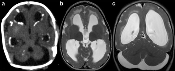
Cerebellar abnormalities are encountered in a high number of neurological diseases that present in the neonatal period. These disorders can be categorized broadly as inherited (e.g. malformations, inborn errors of metabolism) or acquired (e.g. hemorrhages, infections, stroke). In some disorders such as Dandy-Walker malformation or Joubert syndrome, the main abnormalities are located within the cerebellum and brainstem. In other disorders such as Krabbe disease or sulfite oxidase deficiency, the main abnormalities are found within the supratentorial brain, but the cerebellar involvement may be helpful for diagnostic purposes. In In this article, we review neurological disorders with onset in the neonatal period and cerebellar involvement with a focus on how characterization of cerebellar involvement can facilitate accurate diagnosis and improved accuracy of neuro-functional prognosis.
Keywords: Cerebellum; Neonatal neurology; Neonate; Neuroimaging.
Figures
Similar articles
-
Isolated Unilateral Cerebellar Hemispheric Dysplasia: A Rare Entity.Can J Neurol Sci. 2019 Nov;46(6):760-761. doi: 10.1017/cjn.2019.249. Can J Neurol Sci. 2019. PMID: 31352912
-
Cerebellar hypoplasia: differential diagnosis and diagnostic approach.Am J Med Genet C Semin Med Genet. 2014 Jun;166C(2):211-26. doi: 10.1002/ajmg.c.31398. Epub 2014 May 16. Am J Med Genet C Semin Med Genet. 2014. PMID: 24839100
-
Pre- and Postnatal Neuroimaging of Congenital Cerebellar Abnormalities.Cerebellum. 2016 Feb;15(1):5-9. doi: 10.1007/s12311-015-0699-z. Cerebellum. 2016. PMID: 26166429 Review.
-
"Acquired" Dandy-Walker malformation and cerebellar hemorrhage: Usefulness of serial MRI.Eur J Paediatr Neurol. 2016 Jan;20(1):188-91. doi: 10.1016/j.ejpn.2015.09.009. Epub 2015 Oct 9. Eur J Paediatr Neurol. 2016. PMID: 26507178
-
Cerebellar and Brainstem Malformations.Neuroimaging Clin N Am. 2016 Aug;26(3):341-57. doi: 10.1016/j.nic.2016.03.005. Neuroimaging Clin N Am. 2016. PMID: 27423798 Review.
Cited by
-
Physiotherapy and Rehabilitation in a Child with Joubert Syndrome.Case Rep Pediatr. 2017;2017:8076494. doi: 10.1155/2017/8076494. Epub 2017 Aug 23. Case Rep Pediatr. 2017. PMID: 29138705 Free PMC article.
-
Psychosis and Dandy-Walker syndrome: a case report and review of the literature.Gen Psychiatr. 2021 Apr 14;34(2):e100254. doi: 10.1136/gpsych-2020-100254. eCollection 2021. Gen Psychiatr. 2021. PMID: 33937630 Free PMC article.
-
Cerebellar injury and impaired function in a rabbit model of maternal inflammation induced neonatal brain injury.Neurobiol Learn Mem. 2019 Nov;165:106901. doi: 10.1016/j.nlm.2018.07.005. Epub 2018 Jul 24. Neurobiol Learn Mem. 2019. PMID: 30016703 Free PMC article.
-
A Proposed Clinical Classification and a Diagnostic Approach for Congenital Ataxias.Neurol Clin Pract. 2021 Jun;11(3):e328-e336. doi: 10.1212/CPJ.0000000000000966. Neurol Clin Pract. 2021. PMID: 34484907 Free PMC article. Review.
-
Tic-Related Obsessive-Compulsive and Eating Disorders in Dandy-Walker Variant: A Case Report and Systematic Reappraisal of Psychiatric Profiles.Brain Sci. 2024 Apr 6;14(4):362. doi: 10.3390/brainsci14040362. Brain Sci. 2024. PMID: 38672014 Free PMC article. Review.
References
Publication types
Grants and funding
LinkOut - more resources
Full Text Sources
Other Literature Sources
