. 2016 Jan 15;11(1):e0145084.
doi: 10.1371/journal.pone.0145084. eCollection 2016.

Cytoskeletal Expression and Remodeling in Pluripotent Stem Cells

Affiliations

Cytoskeletal Expression and Remodeling in Pluripotent Stem Cells

Liana C Boraas et al. PLoS One. .

Abstract

Many emerging cell-based therapies are based on pluripotent stem cells, though complete understanding of the properties of these cells is lacking. In these cells, much is still unknown about the cytoskeletal network, which governs the mechanoresponse. The objective of this study was to determine the cytoskeletal state in undifferentiated pluripotent stem cells and remodeling with differentiation. Mouse embryonic stem cells (ESCs) and reprogrammed induced pluripotent stem cells (iPSCs), as well as the original un-reprogrammed embryonic fibroblasts (MEFs), were evaluated for expression of cytoskeletal markers. We found that pluripotent stem cells overall have a less developed cytoskeleton compared to fibroblasts. Gene and protein expression of smooth muscle cell actin, vimentin, lamin A, and nestin were markedly lower for ESCs than MEFs. Whereas, iPSC samples were heterogeneous with most cells expressing patterns of cytoskeletal proteins similar to ESCs with a small subpopulation similar to MEFs. This indicates that dedifferentiation during reprogramming is associated with cytoskeletal remodeling to a less developed state. In differentiation studies, it was found that shear stress-mediated differentiation resulted in an increase in expression of cytoskeletal intermediate filaments in ESCs, but not in iPSC samples. In the embryoid body model of spontaneous differentiation of pluripotent stem cells, however, both ESCs and iPSCs had similar gene expression for cytoskeletal proteins during early differentiation. With further differentiation, however, gene levels were significantly higher for iPSCs compared to ESCs. These results indicate that reprogrammed iPSCs more readily reacquire cytoskeletal proteins compared to the ESCs that need to form the network de novo. The strategic selection of the parental phenotype is thus critical not only in the context of reprogramming but also the ultimate functionality of the iPSC-differentiated cell population. Overall, this increased characterization of the cytoskeleton in pluripotent stem cells will allow for the better understanding and design of stem cell-based therapies.


Conflict of interest statement

Competing Interests: The authors have declared that no competing interests exist.

Figures

Fig 1
Fig 1. Morphology of ESCs, iPSCs, and MEFs.
Representative phase images are shown at both low and high magnification. High magnification images of ESCs show a single highly refractive colony and of MEFs show an adherent spread morphology. A low magnification image of iPSCs shows population heterogeneity, including half dome colonies that were highly refractive (single arrow head; high magnification image also shown), colonies that were less tightly compact with a less refractive outer edge (double headed arrows), and clusters of adherent cells (star). All scale bars represent 200 μm.
Fig 2
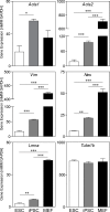
Fig 2. Cytoskeletal gene expression in ESCs, iPSCs, and MEFs.
Cytoskeletal gene expression of microfilaments (Acta1 and Acta2), microtubules (Tuba1b), and both cytoplasmic (Vim and Nes) and nuclear (Lmna) intermediate filaments are shown (all normalized to Gapdh). Data presented are mean ± SEM (n = 3), with significant differences indicated using asterisks (* for p<0.05, ** for p<0.01, and *** for p<0.001).
Fig 3
Fig 3. Immunofluorescent staining for cytoskeletal proteins in ESCs, iPSCs, and MEFs.
Samples were stained for ACTIN (A), LAMIN A/C (B), NESTIN (C) and VIMENTIN (D) (cytoskeletal elements in green) with a nuclear counterstain (blue). Shown are images at low magnification (merged: left columns) and high magnification (merged: middle columns; cytoskeleton only: right columns). Single headed arrows and inserts highlight a subpopulation of cells with a spread morphology in iPSC cultures. Scale bars = 30 μm unless otherwise specified.
Fig 4
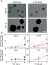
Fig 4. Morphology and cytoskeletal gene expression of differentiating ESCs and iPSCs in the EB model.
(A) Phase images at the same magnification for ESC-EBs and iPSC-EBs at Day 4 and 8. Scale bar represents 200 μm. (B) Gene expression at Day 2, 4, and 8 of Acta1, Vim, Nes, and Lmna (all normalized to Gapdh) of ESC-EBs (red squares) and iPSC-EBs (black circles). Data are presented as mean ± SEM (n = 3), where significant differences are indicated with asterisks (*p<0.05, **p<0.01). For reference, mean values of expression for undifferentiated ESCs (dotted red line) and iPSCs (dashed black line) from Fig 2 are indicated.
Fig 5
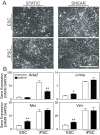
Fig 5. Morphology and cytoskeletal gene expression of ESCs and iPSCs after shear-mediated differentiation.
Evaluation of ESCs and iPSCs after SHEAR treatment or as STATIC controls. (A) Phase images of all four groups. Scale bar represents 200 μm. (B) Gene expression levels for Acta1, Vim, Nes, and Lmna (all normalized to Gapdh) for STATIC (white bars) and SHEAR (black bars) samples. Data are presented as mean ± SEM (n = 5 for ESCs and n = 3 for iPSCs), where significant differences are indicated with asterisks (*p<0.05, **p<0.01).

References

    1. Kim K, Doi A, Wen B, Ng K, Zhao R, Cahan P, et al. (2010) Epigenetic memory in induced pluripotent stem cells. Nature 467: 285–290. 10.1038/nature09342 - DOI - PMC - PubMed
    1. Takahashi K, Yamanaka S (2006) Induction of pluripotent stem cells from mouse embryonic and adult fibroblast cultures by defined factors. Cell 126: 663–676. - PubMed
    1. Takahashi K, Tanabe K, Ohnuki M, Narita M, Ichisaka T, Tomoda K, et al. (2007) Induction of pluripotent stem cells from adult human fibroblasts by defined factors. Cell 131: 861–872. - PubMed
    1. Yu J, Vodyanik MA, Smuga-Otto K, Antosiewicz-Bourget J, Frane JL, Tian S, et al. (2007) Induced pluripotent stem cell lines derived from human somatic cells. Science 318: 1917–1920. - PubMed
    1. Bar-Nur O, Russ HA, Efrat S, Benvenisty N (2011) Epigenetic memory and preferential lineage-specific differentiation in induced pluripotent stem cells derived from human pancreatic islet beta cells. Cell stem cell 9: 17–23. 10.1016/j.stem.2011.06.007 - DOI - PubMed

Publication types

LinkOut - more resources