. 2016 Jan 15;11(1):e0147155.
doi: 10.1371/journal.pone.0147155. eCollection 2016.

Differentiation of Human Induced-Pluripotent Stem Cells into Smooth-Muscle Cells: Two Novel Protocols

Affiliations

Differentiation of Human Induced-Pluripotent Stem Cells into Smooth-Muscle Cells: Two Novel Protocols

Libang Yang et al. PLoS One. .

Abstract

Conventional protocols for differentiating human induced-pluripotent stem cells (hiPSCs) into smooth-muscle cells (SMCs) can be inefficient and generally fail to yield cells with a specific SMC phenotype (i.e., contractile or synthetic SMCs). Here, we present two novel hiPSC-SMC differentiation protocols that yield SMCs with predominantly contractile or synthetic phenotypes. Flow cytometry analyses of smooth-muscle actin (SMA) expression indicated that ~45% of the cells obtained with each protocol assumed an SMC phenotype, and that the populations could be purified to ~95% via metabolic selection. Assessments of cellular mRNA and/or protein levels indicated that SMA, myosin heavy chain II, collagen 1, calponin, transgelin, connexin 43, and vimentin expression in the SMCs obtained via the Contractile SMC protocol and in SMCs differentiated via a traditional protocol were similar, while SMCs produced via the Sythetic SMC protocol expressed less calponin, more collagen 1, and more connexin 43. Differences were also observed in functional assessments of the two SMC populations: the two-dimensional surface area of Contractile SMCs declined more extensively (to 12% versus 44% of original size) in response to carbachol treatment, while quantification of cell migration and proliferation were greater in Synthetic SMCs. Collectively, these data demonstrate that our novel differentiation protocols can efficiently generate SMCs from hiPSCs.


Conflict of interest statement

Competing Interests: The authors have declared that no competing interests exist.

Figures

Fig 1
Fig 1. Chronological summary of the hiPSC-SMC differentiation protocols.
hiPSCs and ESCs were cultured in mTeSRTM medium on Matrigel-coated plates, with daily medium changes, until confluent (~2 days); then, differentiation into mesodermal-lineage cells was initiated on Day 0 by culturing the cells with CHIR99021 and BMP-4 in RPMI1640 medium and 2% B27. Differentiation into Synthetic SMCs or Contractile SMCs began on Day 3. Synthetic SMCs were produced by culturing the cells with VEGF-A and FGFβ in RPMI1640 medium and 2% B27 minus insulin (B27) from Day 3 to Day 7, with VEGF-A and FGFβ in RPMI1640 and 2% B27 (with insulin) from Day 7 to Day 10, and with PDGFβ and TGFβ in RPMI1640 and 2% B27 from Day 10 to Day 14. Contractile SMCs were produced by culturing the cells with VEGF-A and FGFβ in RPMI1640 and 2% B27 from Day 3 to Day 7, and with PDGFβ and TGFβ in RPMI1640 and 2% B27 from Day 7 to Day 14. Purification was performed by maintaining the differentiated cells in 4 mM lactate RPMI1640 metabolic medium for 4 to 6 days.
Fig 2
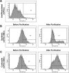
Fig 2. Efficiency of hiPSC-SMC differentiation.
The efficiency of the differentiation protocols was evaluated via flow cytometry analyses of SMA expression in (A) undifferentiated cardiac-lineage hiPSCs (chiPSCs) and in (B-C) chiPSC-SMCs obtained via the (B) Synthetic and (C) Contractile SMC differentiation protocols before and after purification.
Fig 3
Fig 3. hiPSC-SMC marker expression.
(A) mRNA levels of the SMC markers alpha smooth-muscle actin 2 (αSMA-2), smooth muscle myosin heavy chain 11 (MHC-11), calponin (Calp), vascular-endothelial cadherin (VE-Cad), and transgelin (Tgln) were evaluated via quantitative RT-PCR and normalized to endogenous GAPDH mRNA levels (*p<0.01 vs Conventional or Contractile chiPSC-SMCs, p<0.05 vs Conventional chiPSC-SMCs, p<0.01 vs Contractile chiPSC-SMCs). (B) Smooth-muscle actin (SMA), collagen I (Col I), connexin 43 (Cnx 43), vimentin (Vmt), and calponin (Calp) protein expression (red) was detected via immunofluorescent staining in human aortic SMCs, in chiPSC-SMCs that were obtained via a conventional differentiation protocol, and in chiPSC-SMCs obtained via our Synthetic or Contractile hiPSC-SMC differentiation protocols; nuclei were counterstained with DAPI (blue) (bar = 100 μm).
Fig 4
Fig 4. hiPSC-SMC functional assessments.
(A) 4×10⁵ Synthetic or Contractile chiPSC-SMCs were cultured on gelatin-coated plates for 24 hours; then, the plate was scratched with a 200-μL pipette tip, and images of the scratched area were obtained 0 and 10 hours later. Migration was quantified by counting the number of cells that had migrated into the scratched area (*p<0.01). (B) 1×10⁶/mL Synthetic or Contractile chiPSC-SMCs were suspended in 100 μL of RPMI1640 and cultured in the presence of PDGFβ or TGFβ for 90 min; then, the solutions were serially diluted in half six times, and cell concentrations were evaluated via optical density measurements at 490 nm (*p<0.01). (C) 2×10⁵ Synthetic or Contractile chiPSC-SMCs were cultured on gelatin-coated plates for 24 hours; then, the cells were treated with carbachol to induce contraction, and images were obtained 0 and 5 min later. (D) Contraction was evaluated by calculating the mean cell surface area at each of the two time points (*p<0.01). (E) 1×10⁶ Synthetic or Contractile chiPSC-SMCs were suspended in a fibrinogen gel; then, the gels were cultured with aprotinin and Rho kinase inhibitor, and the surface area of the gels was measured 0 and 3 days later (*p<0.01).

References

    1. Zhang S, Dutton JR, Su L, Zhang J, Ye L. The influence of a spatiotemporal 3D environment on endothelial cell differentiation of human induced pluripotent stem cells. Biomaterials 2014;35(12):3786–93. 10.1016/j.biomaterials.2014.01.037 - DOI - PMC - PubMed
    1. Ye L, Chang YH, Xiong Q, Zhang P, Zhang L, Somasundaram P, et al. Cardiac repair in a porcine model of acute myocardial infarction with human induced pluripotent stem cell-derived cardiovascular cells. Cell Stem Cell 2014;15(6):750–61. 10.1016/j.stem.2014.11.009 - DOI - PMC - PubMed
    1. Marchand M, Anderson EK, Phadnis SM, Longaker MT, Cooke JP, Chen B, et al. Concurrent generation of functional smooth muscle and endothelial cells via a vascular progenitor. Stem Cells Transl Med 2014;3(1):91–7. 10.5966/sctm.2013-0124 - DOI - PMC - PubMed
    1. Dash BC, Jiang Z, Suh C, Qyang Y. Induced pluripotent stem cell-derived vascular smooth muscle cells: methods and application. Biochem J 2015;465(2):185–94. 10.1042/BJ20141078 - DOI - PMC - PubMed
    1. Jiang X, Rowitch DH, Soriano P, McMahon AP, Sucov HM. Fate of the mammalian cardiac neural crest. Development 2000;127(8):1607–16. - PubMed

Publication types

MeSH terms