. 2016 Jan 13;17(1):96.
doi: 10.3390/ijms17010096.

Overexpression of PRL7D1 in Leydig Cells Causes Male Reproductive Dysfunction in Mice

Affiliations

Overexpression of PRL7D1 in Leydig Cells Causes Male Reproductive Dysfunction in Mice

Yaping Liu et al. Int J Mol Sci. .

Abstract

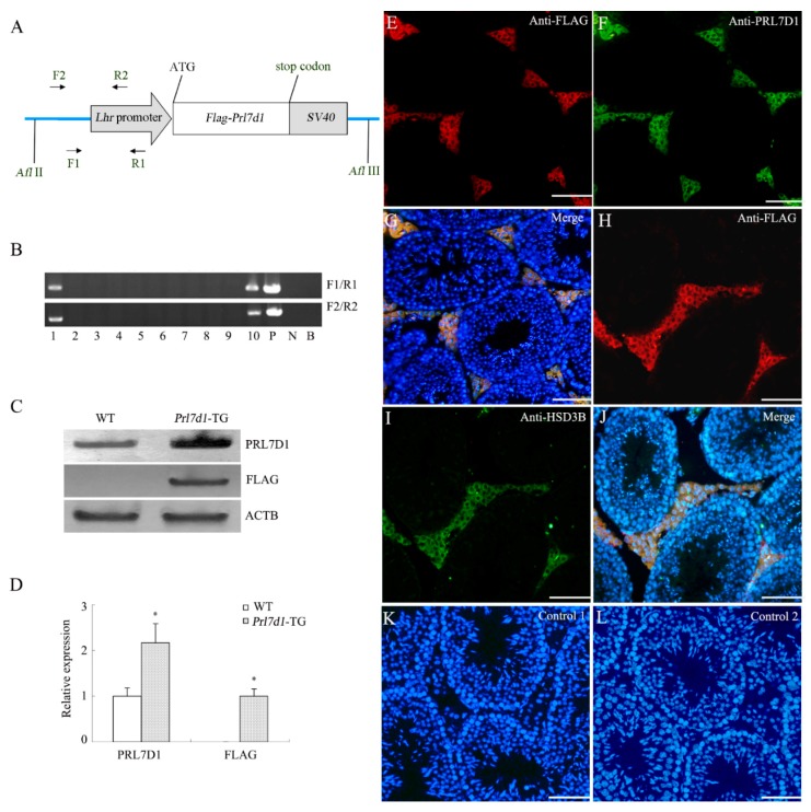
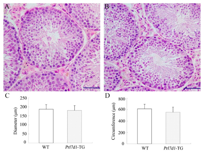
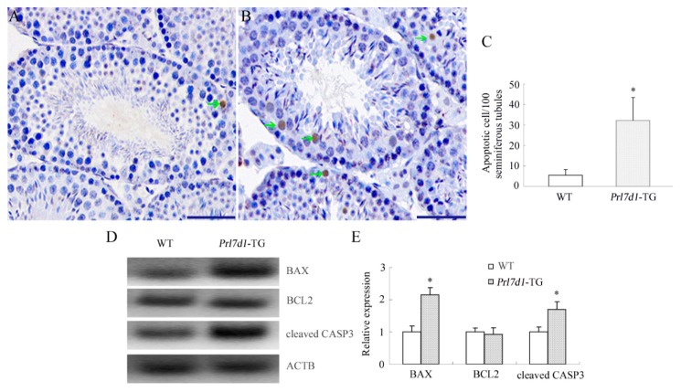
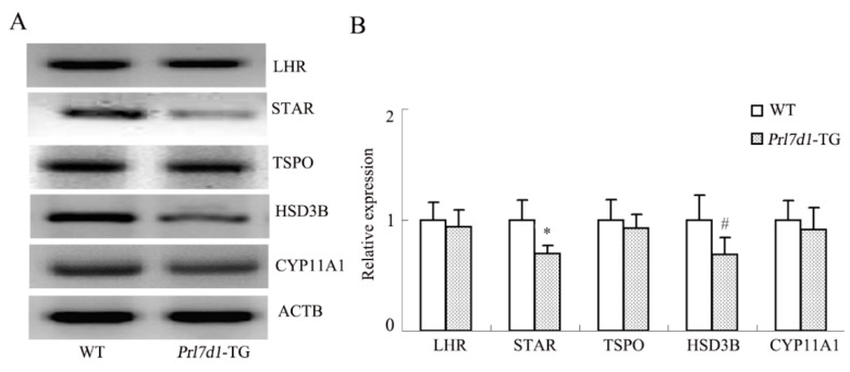
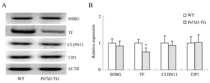
Prolactin family 7, subfamily d, member 1 (PRL7D1) is found in mouse placenta. Our recent work showed that PRL7D1 is also present in mouse testis Leydig cells, and the expression of PRL7D1 in the testis exhibits an age-related increase. In the present study, we generated transgenic mice with Leydig cell-specific PRL7D1 overexpression to explore its function during male reproduction. Prl7d1 male mice exhibited subfertility as reflected by reduced sperm counts and litter sizes. The testes from Prl7d1 transgenic mice appeared histologically normal, but the frequency of apoptotic germ cells was increased. Prl7d1 transgenic mice also had lower testosterone concentrations than wild-type mice. Mechanistic studies revealed that Prl7d1 transgenic mice have defects in the testicular expression of steroidogenic acute regulatory protein (STAR) and hydroxy-delta-5-steroid dehydrogenase, 3 beta- and steroid delta-isomerase cluster (HSD3B). Further studies revealed that PRL7D1 overexpression affected the expression of transferrin (TF) in Sertoli cells. These results suggest that PRL7D1 overexpression could lead to increased germ cell apoptosis and exert an inhibitory effect on testosterone production in Leydig cells by reducing the expression of certain steroidogenic-related genes. In addition, PRL7D1 appears to have important roles in the function of Sertoli cells, which, in turn, affects male fertility. We conclude that the expression level of PRL7D1 is associated with the reproductive function of male mice.

Keywords: Leydig cells; Plr7d1; male reproductive dysfunction.


Figures

Figure 1
Figure 1
Generation of transgenic mice overexpressing Prl7d1. (A) Schematic representation of the Prl7d1 transgenic construct. The Lhr promoter was fused to Flag-tagged mouse Prl7d1 cDNA and SV40 polyadenylase signal. The FLAG epitope (DYKDDDDK) was introduced into the amino-terminus of the PRL7D1 protein. The primers used for the detection of the transgene are indicated by arrows; (B) PCR analysis of the genomic integration of the mouse Prl7d1 transgene. N, negative control; P, positive control (Prl7d1 transgenic construct); B, blank control; (C) Western blot analyses of PRL7D1, FLAG and actin beta (ACTB) in the testes of four-month-old Prl7d1 transgenic (Prl7d1-TG) and wild-type (WT) mice; (D) The relative expression of PRL7D1 and the FLAG-tagged protein in the testes of mice are shown following their normalization against ACTB. Data are expressed as the means ± SD (n = 6). The protein level within the WT group was set to one. * p < 0.01 compared to the WT group; (EL) Double immunofluorescence staining revealed that FLAG-tagged protein (red fluorescence) co-localized with PRL7D1 (green fluorescence) (EG) or HSD3B (green fluorescence) (HJ) in Leydig cells of adult Prl7d1 mouse testes. Control 1: testicular tissue sections were incubated with IgG1; Control 2: immunofluorescent staining of FLAG-tagged protein in WT testis sections. There was no positive staining in Leydig cells. Staining with DAPI (blue fluorescence) was used to label the nuclei of individual cells found in the section. The scale bar is 50 μm.
Figure 2
Figure 2
Histological analyses of testis from four-month-old wild-type (WT) (A) and Prl7d1 transgenic (Prl7d1-TG) mice (B). Sections were stained with hematoxylin and eosin. The scale bar is 50 μm; (C) The diameter of the seminiferous tubules and (D) the circumference of the seminiferous tubules were normal in Prl7d1 transgenic mice. Results for each measurement are expressed as the means ± SD from a minimum of 60 randomly-selected seminiferous tubules from six mice of each phenotype.
Figure 3
Figure 3
PRL7D1 overexpression results in increased germ cell apoptosis and upregulated expression of cleaved CASP3 and BAX in testes. Representative microphotographs of the TUNEL assay performed in testis sections from wild-type (WT) (A) and Prl7d1 transgenic (Prl7d1-TG) (B) mice. TUNEL-positive cells are stained brown (green arrows). The scale bar is 50 μm; (C) Quantitative assessment of the TUNEL-positive cell number per 100 tubules/testis (n = 6); (D,E) Protein levels of BAX, BCL2 and cleaved CASP3 in testes were detected by Western blot analyses. The relative expression levels of the proteins were determined using densitometric analyses of the data that were normalized against the ACTB signal (n = 6). The protein level in the WT group was set to one. Each bar represents the mean ± SD. * p < 0.01 compared to the WT group.
Figure 4
Figure 4
The levels of serum testosterone (A), LH (C) and GnRH (D) and intra-testicular testosterone (B) in four-month-old Prl7d1 transgenic (Prl7d1-TG) and wild-type (WT) mice. Testosterone, LH and GnRH levels were measured by ELISA. Data are expressed as the means ± SD (n = 6). * p < 0.01 compared to the WT group.
Figure 5
Figure 5
Western blot analyses of the LHR, STAR, TSPO, HSD3B and CYP11A1 in the testes of wild-type (WT) and Prl7d1 transgenic (Prl7d1-TG) mice. (A) Representative Western immunoblots; (B) The relative expression levels of the proteins were determined using densitometric analyses of the data that were normalized against the ACTB signal (n = 6). The protein level in the control group was set to one. Each bar represents the mean ± SD. # p < 0.05, * p < 0.01 compared to the WT group.
Figure 6
Figure 6
Western blot analyses of the SHBG, TF, CLDN11 and TJP1 in the testes of wild-type (WT) and Prl7d1 transgenic (Prl7d1-TG) mice. (A) Representative western immunoblots; (B) The relative expression levels of the proteins were determined using densitometric analyses of the data that were normalized against the ACTB signal (n = 6). The protein level in the control group was set to one. Each bar represents the mean ± SD. * p < 0.01 compared to the WT group.

Similar articles

References

    1. Soares M.J. The prolactin and growth hormone families: Pregnancy-specific hormones/cytokines at the maternal-fetal interface. Reprod. Biol. Endocrinol. 2004;2:51. doi: 10.1186/1477-7827-2-51. - DOI - PMC - PubMed
    1. Alam S.M., Ain R., Konno T., Ho-Chen J.K., Soares M.J. The rat prolactin gene family locus: Species-specific gene family expansion. Mamm. Genome. 2006;17:858–877. doi: 10.1007/s00335-006-0010-1. - DOI - PubMed
    1. Green J.A. Defining the function of a prolactin gene family member. Proc. Natl. Acad. Sci. USA. 2004;101:16397–16398. doi: 10.1073/pnas.0406934101. - DOI - PMC - PubMed
    1. Bachelot A., Binart N. Reproductive role of prolactin. Reproduction. 2007;133:361–369. doi: 10.1530/REP-06-0299. - DOI - PubMed
    1. Haig D. Placental growth hormone-related proteins and prolactin-related proteins. Placenta. 2008;29(Suppl. A):S36–S41. doi: 10.1016/j.placenta.2007.09.010. - DOI - PubMed

Publication types

MeSH terms

LinkOut - more resources