Review
. 2016 Jan 12;5(1):7.
doi: 10.3390/jcm5010007.

Reversible Human TGF-β Signal Shifting between Tumor Suppression and Fibro-Carcinogenesis: Implications of Smad Phospho-Isoforms for Hepatic Epithelial-Mesenchymal Transitions

Affiliations
Review

Reversible Human TGF-β Signal Shifting between Tumor Suppression and Fibro-Carcinogenesis: Implications of Smad Phospho-Isoforms for Hepatic Epithelial-Mesenchymal Transitions

Katsunori Yoshida et al. J Clin Med. .

Abstract

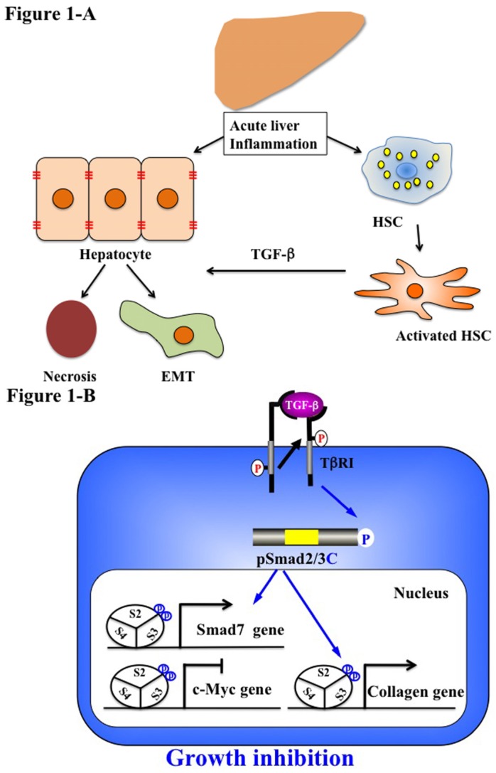
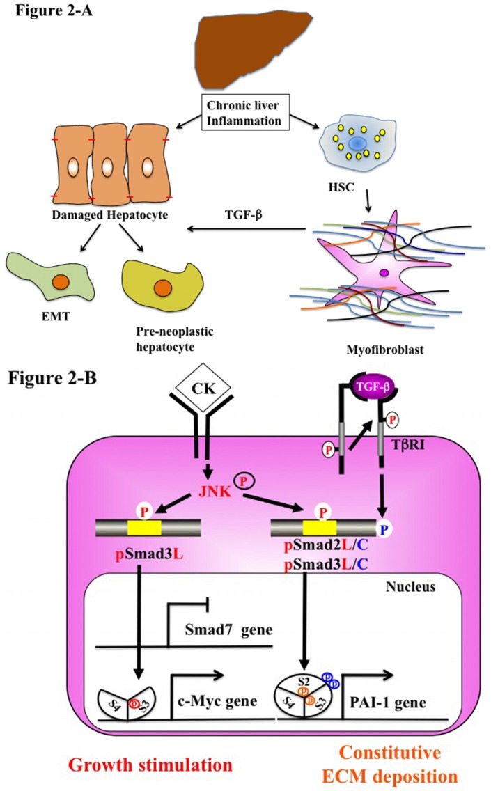
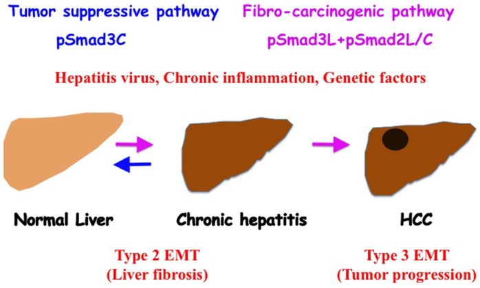
Epithelial-mesenchymal transition (EMT) and mesenchymal-epithelial transition (MET) are observed during both physiological liver wound healing and the pathological fibrotic/carcinogenic (fibro-carcinogenetic) process. TGF-β and pro-inflammatory cytokine are considered to be the major factors accelerating liver fibrosis and promoting liver carcinogenesis. Smads, consisting of intermediate linker regions connecting Mad homology domains, act as the intracellular mediators of the TGF-β signal transduction pathway. As the TGF-β receptors, c-Jun N-terminal kinase and cyclin-dependent kinase, differentially phosphorylate Smad2/3, we have generated numerous antibodies against linker (L) and C-terminal (C) phosphorylation sites in Smad2/3 and identified four types of phosphorylated forms: cytostatic COOH-terminally-phosphorylated Smad3 (pSmad3C), mitogenic pSmad3L (Ser-213) signaling, fibrogenic pSmad2L (Ser-245/250/255)/C signaling and migratory pSmad2/3L (Thr-220/179)/C signaling. After acute liver injury, TGF-β upregulates pSmad3C signaling and terminates pSmad3L (Ser-213)-mediated hepatocyte proliferation. TGF-β and pro-inflammatory cytokines cooperatively enhance collagen synthesis by upregulating pSmad2L (Thr-220)/C and pSmad3L (Thr-179)/C pathways in activated hepatic stellate cells. During chronic liver injuries, hepatocytes persistently affected by TGF-β and pro-inflammatory cytokines eventually become pre-neoplastic hepatocytes. Both myofibroblasts and pre-neoplastic hepatocyte exhibit the same carcinogenic (mitogenic) pSmad3L (Ser-213) and fibrogenic pSmad2L (Ser-245/250/255)/C signaling, with acquisition of fibro-carcinogenic properties and increasing risk of hepatocellular carcinoma (HCC). Firstly, we review phospho-Smad-isoform signalings in epithelial and mesenchymal cells in physiological and pathological conditions and then consider Smad linker phosphorylation as a potential target for pathological EMT during human fibro-carcinogenesis, because human Smad phospho-isoform signals can reverse from fibro-carcinogenesis to tumor-suppression in a process of MET after therapy.

Keywords: Smad; epithelial-mesenchymal transition (EMT); hepatic stellate cells (HSC); liver fibro-carcinogenesis; myofibroblast (MFB); transforming growth factor-β (TGF-β).


Figures

Figure 1
Figure 1
Liver regeneration-related EMT and phospho-Smad signaling in acute liver disease. (A) Quiescent hepatic stellate cells (HSC) are characterized by retinoid droplets in the cytoplasm. Acute liver injury caused HSC activation and hepatocyte damage, necrosis and EMT. Activated HSC move from the space of Disse to sites of damage where the activated HSC contribute to tissue repair by producing large amounts of collagen. HSC also play an important role in secreting TGF-β. (B) Catalytically-active TβRI phosphorylates COOH-tail serine residues of Smad2 and Smad3. Both pSmad2C and pSmad3C are localized in the nuclei of hepatocytes and mesenchymal cells in acute injured liver. After binding with Smad4, pSmad2/3C translocate with Smad4 to the nucleus and bind to the collagen promoter. pSmad2/3C stimulate extracellular matrix (ECM) deposition and suppress cell growth by c-Myc inhibition. However, Smad7 induced by the pSmad3L/C signal terminates the fibrogenic phospho-Smad signaling. This negative feedback mechanism of the fibrogenic TGF-β/CK signal results in a transient collagen synthesis in the activated HSC, which may thus contribute to tissue repair.
Figure 2
Figure 2
Liver fibro-carcinogenesis-related EMT and phospho-Smad signaling during chronic liver disease. (A) Prolonged exposure to chronic injury; HSC undergo constitutive activation to become myofibroblasts (MFB)-like cells, which persistently induce deposition of ECM and liver fibrosis. Continuous insults will shift EMT-like cells to complete EMT and pre-neoplastic hepatocytes. (B) During chronic liver injury, pro-inflammatory cytokines (CK), such as TNF-α activate JNK, result in phosphorylation of both Smad2L and Smad3L, both in MFB and pre-neoplastic hepatocyte. P-Smad3L translocates with Smad4 to the nucleus and binds the PAI-1 promoter. After COOH-tail phosphorylation of cytoplasmic pSmad2L by TβRI, pSmad2L/C translocates to the nucleus. Both pSmad2L/C and pSmad3L stimulates PAI-1 transcription and ECM deposition, while they suppress the pSmad3C-mediated tumor suppressive pathway. Pre-neoplastic hepatocytes exhibit the same oncogenic (mitogenic) pSmad3L and fibrogenic pSmad2L signaling as MFB, thereby accelerating liver fibrosis and increasing the risk of HCC. In contrast to Smad7 induction in HSC via the pSmad3C pathway, pSmad3L cannot induce Smad7 in MFB and pre-neoplastic hepatocytes (left). Under a low level of Smad7, the fibrogenic phospho-Smad signaling can constitutively promote ECM deposition by MFB, which may eventually develop into accelerated liver fibro-carcinogenesis.
Figure 3
Figure 3
Phosphorylated Smad2/3 signaling fibro-carcinogenesis. As human hepatitis virus-related chronic liver diseases progress, chronic inflammation and hepatitis virus additively shift hepatocytic Smad phospho-isoform signaling from tumor-suppressive pSmad3C to the fibro-carcinogenic pSmad3L and pSmad2L/C pathway. Anti-viral therapy can reverse phospho-Smad signaling from fibro-carcinogenesis to tumor suppression. Type 2 EMT promotes liver fibrosis induced by chronic inflammation. Type 3 EMT exacerbates the HCC phenotype by upregulating invasive and metastatic potential.

References

    1. Greenburg G., Hay E.D. Epithelia suspended in collagen gels can lose polarity and express characteristics of migrating mesenchymal cells. J. Cell Biol. 1982;95:333–339. doi: 10.1083/jcb.95.1.333. - DOI - PMC - PubMed
    1. Kalluri R., Neilson E.G. Epithelial-mesenchymal transition and its implications for fibrosis. J. Clin. Investig. 2003;112:1776–1784. doi: 10.1172/JCI200320530. - DOI - PMC - PubMed
    1. Kalluri R., Weinberg R.A. The basics of epithelial-mesenchymal transition. J. Clin. Investig. 2009;119:1420–1428. doi: 10.1172/JCI39104. - DOI - PMC - PubMed
    1. Zavadil J., Bottinger E.P. TGF-β and epithelial-to-mesenchymal transitions. Oncogene. 2005;24:5764–5774. doi: 10.1038/sj.onc.1208927. - DOI - PubMed
    1. Moses H.L., Serra R. Regulation of differentiation by TGF-β. Curr. Opin. Genet. Dev. 1996;6:581–586. doi: 10.1016/S0959-437X(96)80087-6. - DOI - PubMed

LinkOut - more resources