Gut microbiota lipopolysaccharide accelerates inflamm-aging in mice
- PMID: 26772806
- PMCID: PMC4715324
- DOI: 10.1186/s12866-016-0625-7
Gut microbiota lipopolysaccharide accelerates inflamm-aging in mice
Abstract
Background: The constitutive inflammation that characterizes advanced age is termed inflamm-aging. This process is associated with age-related changes to immune homeostasis and gut microbiota. We investigated the relationship between aging and gut microbiota lipopolysaccharide (LPS)-inducible inflammation.
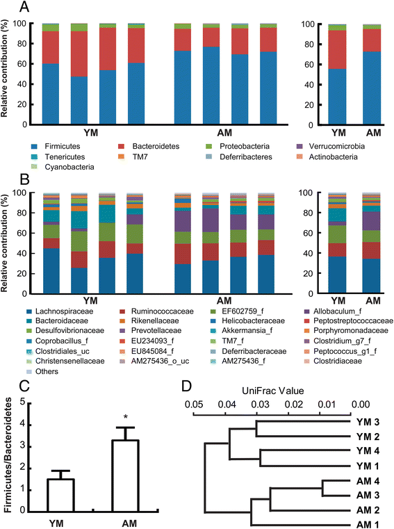
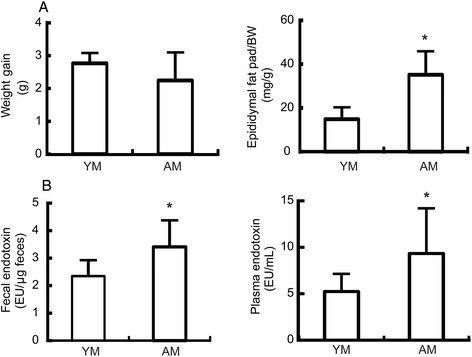
Results: A taxonomy-based analysis showed that aging resulted in increased prevalence of the phyla Firmicutes and Actinobacteria and a reduced prevalence of Bacteroidetes and Tenericutes, resulting in an increase in the Firmicutes to Bacteroidetes ratio. The levels of plasmatic and fecal lipopolysaccharides were higher in aged mice. Aging induced the expression of p16 and the activation of nuclear factor-kappa B (NF-κB) in the colon of aged mice. Interestingly, the expression level of sterile α-motif domain- and HD domain-containing protein 1 (SAMHD1) in the colon was higher in aged mice than in young mice, while cyclin-dependent kinase-2 and cyclin E levels were lower in aged mice than in young mice. The lipopolysaccharide fraction of fecal lysates (LFL) from young or aged mice increased p16 and SAMHD1 expression and NF-κB activation in peritoneal macrophages from wild-type mice, in a TLR4-dependent manner. However, LFLs did not induce NF-κB activation and SAMHD1 expression in peritoneal macrophages from TLR4-deificent mice, whereas they significantly induced p16 expression. Nevertheless, p16 expression was induced more potently in macrophages from WT mice than in macrophages from TLR4-deficient mice.
Conclusion: Aging increased p16 and SAMHD1 expression, gut microbiota LPS production, and NF-κB activation; thereby, signifying that gut microbiota LPS may accelerate inflamm-aging and SAMHD1 may be an inflamm-aging marker.
Figures


Similar articles
-
Lactobacillus rhamnosus HDB1258 modulates gut microbiota-mediated immune response in mice with or without lipopolysaccharide-induced systemic inflammation.BMC Microbiol. 2021 May 13;21(1):146. doi: 10.1186/s12866-021-02192-4. BMC Microbiol. 2021. PMID: 33985438 Free PMC article.
-
Orally administrated Lactobacillus pentosus var. plantarum C29 ameliorates age-dependent colitis by inhibiting the nuclear factor-kappa B signaling pathway via the regulation of lipopolysaccharide production by gut microbiota.PLoS One. 2015 Feb 17;10(2):e0116533. doi: 10.1371/journal.pone.0116533. eCollection 2015. PLoS One. 2015. PMID: 25689583 Free PMC article.
-
Bifidobacterium adolescentis IM38 ameliorates high-fat diet-induced colitis in mice by inhibiting NF-κB activation and lipopolysaccharide production by gut microbiota.Nutr Res. 2017 May;41:86-96. doi: 10.1016/j.nutres.2017.04.003. Epub 2017 Apr 19. Nutr Res. 2017. PMID: 28479226
-
Managing the manager: gut microbes, stem cells and metabolism.Diabetes Metab. 2014 Jun;40(3):186-90. doi: 10.1016/j.diabet.2013.12.004. Epub 2014 Jan 23. Diabetes Metab. 2014. PMID: 24462190 Review.
-
Understanding gut microbiota in elderly's health will enable intervention through probiotics.Benef Microbes. 2014 Sep;5(3):235-46. doi: 10.3920/BM2013.0079. Benef Microbes. 2014. PMID: 24889891 Review.
Cited by
-
Metagenomics Study Reveals Changes in Gut Microbiota in Centenarians: A Cohort Study of Hainan Centenarians.Front Microbiol. 2020 Jul 2;11:1474. doi: 10.3389/fmicb.2020.01474. eCollection 2020. Front Microbiol. 2020. PMID: 32714309 Free PMC article.
-
Intestinal microbiota: a new perspective on delaying aging?Front Microbiol. 2023 Nov 30;14:1268142. doi: 10.3389/fmicb.2023.1268142. eCollection 2023. Front Microbiol. 2023. PMID: 38098677 Free PMC article. Review.
-
The influence of gut microbiota alteration on age-related neuroinflammation and cognitive decline.Neural Regen Res. 2022 Nov;17(11):2407-2412. doi: 10.4103/1673-5374.335837. Neural Regen Res. 2022. PMID: 35535879 Free PMC article. Review.
-
Among older adults, age-related changes in the stool microbiome differ by HIV-1 serostatus.EBioMedicine. 2019 Feb;40:583-594. doi: 10.1016/j.ebiom.2019.01.033. Epub 2019 Jan 23. EBioMedicine. 2019. PMID: 30685386 Free PMC article.
-
Aging amplifies a gut microbiota immunogenic signature linked to heightened inflammation.Aging Cell. 2024 Aug;23(8):e14190. doi: 10.1111/acel.14190. Epub 2024 May 9. Aging Cell. 2024. PMID: 38725282 Free PMC article.
References
-
- Mitsuoka T. Bifidobacteria and their role in human health. J Ind Microbiol. 1990;6:263–7. doi: 10.1007/BF01575871. - DOI
Publication types
MeSH terms
Substances
LinkOut - more resources
Full Text Sources
Other Literature Sources
Medical
Research Materials
Miscellaneous

