. 2016 Mar 10;127(10):1276-86.
doi: 10.1182/blood-2014-10-606988. Epub 2016 Jan 14.

B-cell survival and development controlled by the coordination of NF-κB family members RelB and cRel

Affiliations

B-cell survival and development controlled by the coordination of NF-κB family members RelB and cRel

Jonathan V Almaden et al. Blood. .

Abstract

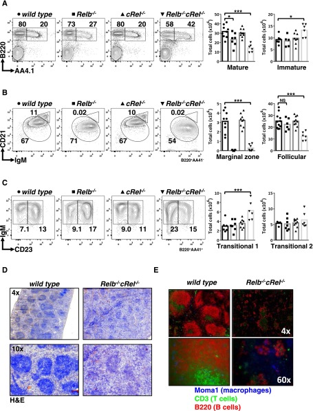
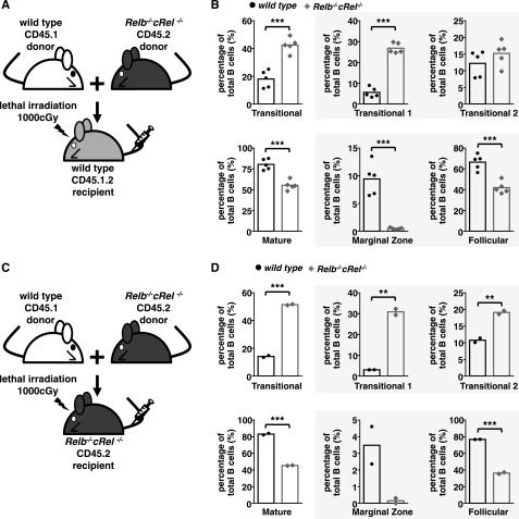
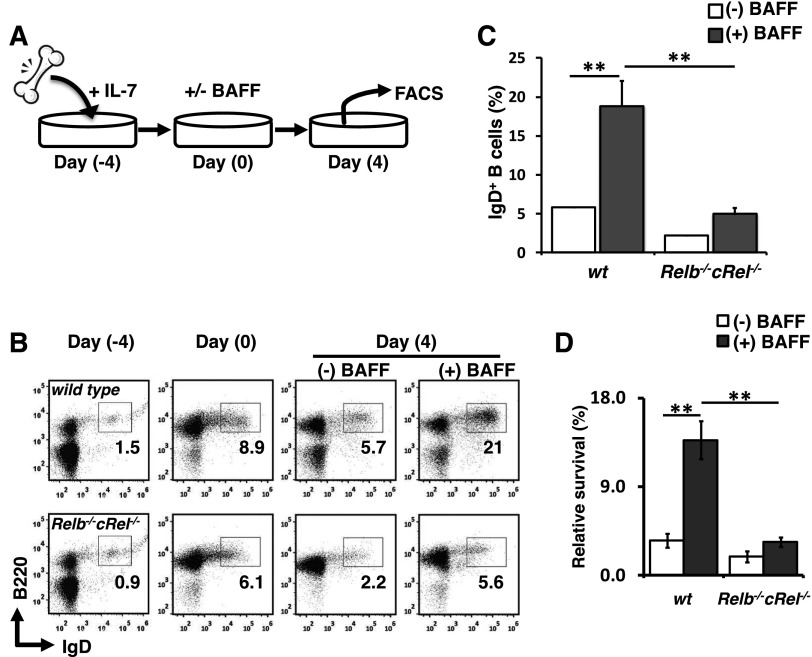
Targeted deletion of BAFF causes severe deficiency of splenic B cells. BAFF-R is commonly thought to signal to nuclear factor κ-light-chain-enhancer of activated B cells (NF-κB)-inducing kinase dependent noncanonical NF-κB RelB. However, RelB-deficient mice have normal B-cell numbers. Recent studies showed that BAFF also signals to the canonical NF-κB pathway, and we found that both RelB and cRel are persistently activated, suggesting BAFF signaling coordinates both pathways to ensure robust B-cell development. Indeed, we report now that combined loss of these 2 NF-κB family members leads to impaired BAFF-mediated survival and development in vitro. Although single deletion of RelB and cRel was dispensable for normal B-cell development, double knockout mice displayed an early B-cell developmental blockade and decreased mature B cells. Despite disorganized splenic architecture in Relb(-/-)cRel(-/-) mice, generation of mixed-mouse chimeras established the developmental phenotype to be B-cell intrinsic. Together, our results indicate that BAFF signals coordinate both RelB and cRel activities to ensure survival during peripheral B-cell maturation.


Figures

Figure 1
Figure 1
BAFF-R stimulation triggers persistent RelB and cRel activity to control in vitro survival functions. (A) BAFF, which binds TACI and BAFF-R, is a key regulator of peripheral B-cell maintenance. BAFF-R signaling functions occur primarily in an NIK-dependent, noncanonical NF-κB manner, mediated through RelB activity, whereas TACI triggers NEMO-dependent canonical NF-κB. (B) Identification of activated NF-κB species in B cells stimulated with BAFF for 24 hours by supershift analysis. (C) RelA, RelB, and cRel EMSA (see “Materials and methods”) of B cells are used to reveal their respective activity kinetics following BAFF-R perturbation. (D) Quantification of RelA, RelB, and cRel activities of BAFF-stimulated primary B cells derived from EMSA. Naive is resting, unstimulated B cells; early and late activity correspond to activities following 1 to 5 hours and 24+ hours of BAFF stimulation, respectively. Representative of ≥4 experiments. *P < .05; **P < .01. (E) Summary table of known NF-κB–deficient mouse models and the effect on B-cell populations. Gray boxes denote a B-cell developmental defect. +++, cell numbers equate to typical wild-type level; ++++, greater than wild-type; ++, intermediate level; +, low; -, not present. EMSA for panels B-C representative of at least 3 experiments.
Figure 2
Figure 2
RelB and cRel coordinate together to provide proper BAFF-mediated survival signals in vitro. (A) FACS plots of in vitro survival assay of whole splenic wild-type, Relb−/−, cRel−/−, and Relb−/−cRel−/− B cells stimulated with BAFF ligand for 70 hours. Numbers represent the percentage of live cells (7AAD) found in culture. Graphical representation of the FACS plots (right), n = 3. (B) FACS plots of in vitro survival assay of FO (CD23+) wild-type and Relb−/−cRel−/− B cells stimulated with BAFF for 40 hours. (C) RNA-seq analysis from BAFF-stimulated CD23+ wild-type, cRel−/−, and Relb−/−cRel−/− B cells at the indicated time points. Genes (517) were upregulated in BAFF-stimulated follicular B cells; 289 of these showed substantial expression defect in B cells lacking both cRel and RelB (middle and bottom panels). Of these, 127 showed expression defects even in the single cRel knockout (middle panel); 162 showed expression defects only in the Relb−/−cRel−/− double knockout (bottom panel). (D) cRel-dependent genes protect cells against cell death. Gene ontology analysis identifies distinct process terms for cRel-dependent vs RelB/cRel-dependent gene clusters. Whereas RelB/cRel-dependent clusters are significantly associated with terms describing metabolic processes, the cRel-dependent cluster shows overrepresentation of negative regulation of cell death/apoptosis. *P < .05; **P < .005; ***P < .001. Also see supplemental Figure 1. (E) List of representative cRel-dependent and RelB/cRel-dependent genes identified as “negative regulators of cell death” and “metabolic process,” respectively, by gene ontology analysis.
Figure 3
Figure 3
Compound deletion of RelB and cRel results in developmental block at the T1 stage. (A-C) FACS analysis of peripheral B-cell development in whole splenic extracts from wild-type, Relb−/−, cRel−/−, and Relb−/−cRel−/− mice. Identification of mature (B220+AA4.1) and transitional B cells (B220+AA4.1+). Mature B cells (B220+AA4.1) are further classified into FO B cells (CD21+IgM+) and mature MZ B cells (CD21highIgM+). Transitional B cells (B220+AA4.1+) are subdivided into T1 B cells (CD23IgM+) and T2 B cells (CD23+IgM+). Scatter plots are graphical representation of FACS plots: wild-type (●), Relb−/− (▪), cRel−/− (▲), and Relb−/−cRel−/− (▼). (D) Histologic analysis of splenic sections taken from wild-type and Relb−/−cRel−/− mouse using hematoxylin and eosin stain (H&E). (E) In vivo analysis of mature B-cell development in double knockout mouse using immunofluorescence of frozen splenic sections stained with anti-CD3 fluorescein isothiocyanate, anti-B220 allophycocyanin, and anti-MOMA-1 Alexa Fluor 405. *P < .05; **P < .005; ***P < .001. Also see supplemental Figure 2.
Figure 4
Figure 4
B-cell developmental defect displayed in Relb−/−cRel−/− mouse is B-cell intrinsic. (A and C) Schematic for the generation of CD45.1 wild-type and CD45.2 Relb−/−cRel−/− mixed bone marrow chimeras. Bone marrow stem cells from CD45.1 wild-type and CD45.2 Relb−/−cRel−/− are mixed in a 50:50 ratio and then injected into a lethally irradiated CD45.1.2 wild-type or Relb−/−cRel−/− mouse. (B and D) Graphical plots of percentages of specific subsets relative to total B cells in indicated mixed bone marrow chimeras, as described previously. The gating strategy used in the analysis is depicted in Figure 3. Representative FACS plots see supplemental Figure 3. For panel B, n = 5; for panel D, n = 2. *P < .05; **P < .005; ***P < .001.
Figure 5
Figure 5
Relb−/−cRel−/− B-cell progenitors fail to respond to BAFF-mediated developmental signal in vitro. (A) Schematic of in vitro B-cell differentiation system. (B) Representative FACS plots of B220 and IgD expression in transitional B cells at the beginning (day 0) and end (day 4) of culture with or without BAFF. (C) Mean frequency of B220+IgD+ (T2-like) B cells following 4 days of culture as described in panel B. Numbers represent frequencies of live B220+ B cells in indicated gates. (D) Survival of in vitro cultured B cells. Live cells were identified by gating out 7AADHi population. Fold survival calculated from initial B-cell population (day 0). For FACS plots, n = 4. wt, wild-type.
Figure 6
Figure 6
Deregulated NF-κB activity in IκBα−/−IκBε−/−cRel−/−TNF−/− leads to reduction in T1 cells and increased MZ B-cell generation. FACS analysis of peripheral B-cell development in whole splenic extracts from wild-type, cRel−/−, Relb−/−cRel−/−, and IκBα−/−IκBε-/cRel−/− TNF−/− mice. The gating strategy used in the analysis is depicted in Figure 3. Numbers represent percentage of denoted subsets as proportion of total B cells. Representative FACS results of 3 experiments shown.

References

    1. Hardy RR, Carmack CE, Shinton SA, Kemp JD, Hayakawa K. Resolution and characterization of pro-B and pre-pro-B cell stages in normal mouse bone marrow. J Exp Med. 1991;173(5):1213–1225. - PMC - PubMed
    1. Hardy RR, Hayakawa K. A developmental switch in B lymphopoiesis. Proc Natl Acad Sci USA. 1991;88(24):11550–11554. - PMC - PubMed
    1. Allman DM, Ferguson SE, Cancro MP. Peripheral B cell maturation. I. Immature peripheral B cells in adults are heat-stable antigenhi and exhibit unique signaling characteristics. J Immunol. 1992;149(8):2533–2540. - PubMed
    1. Allman DM, Ferguson SE, Lentz VM, Cancro MP. Peripheral B cell maturation. II. Heat-stable antigen(hi) splenic B cells are an immature developmental intermediate in the production of long-lived marrow-derived B cells. J Immunol. 1993;151(9):4431–4444. - PubMed
    1. Carsetti R, Köhler G, Lamers MC. Transitional B cells are the target of negative selection in the B cell compartment. J Exp Med. 1995;181(6):2129–2140. - PMC - PubMed

Publication types

MeSH terms

Substances