. 2016 May 5;127(18):2203-13.
doi: 10.1182/blood-2015-09-672352. Epub 2016 Jan 15.

Diffuse large B-cell lymphoma patient-derived xenograft models capture the molecular and biological heterogeneity of the disease

Affiliations

Diffuse large B-cell lymphoma patient-derived xenograft models capture the molecular and biological heterogeneity of the disease

Bjoern Chapuy et al. Blood. .

Abstract

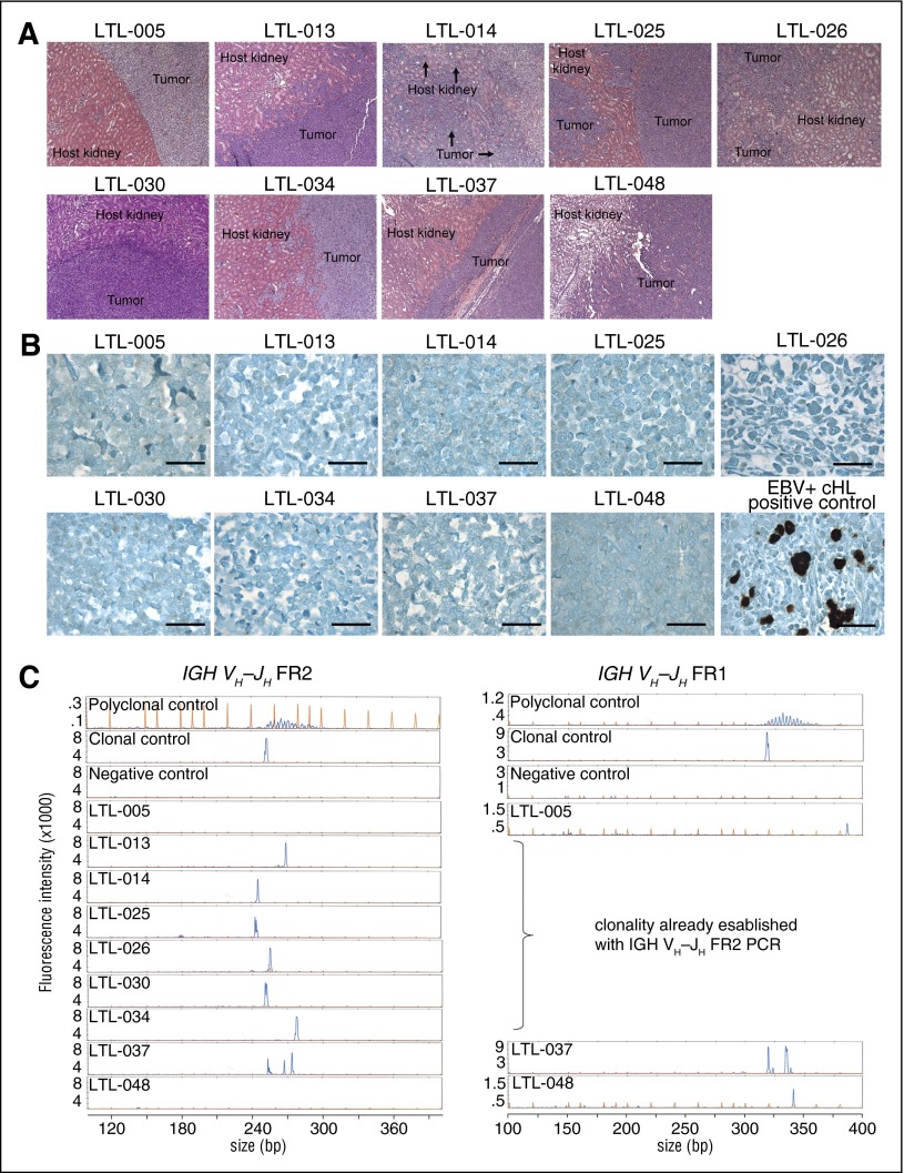
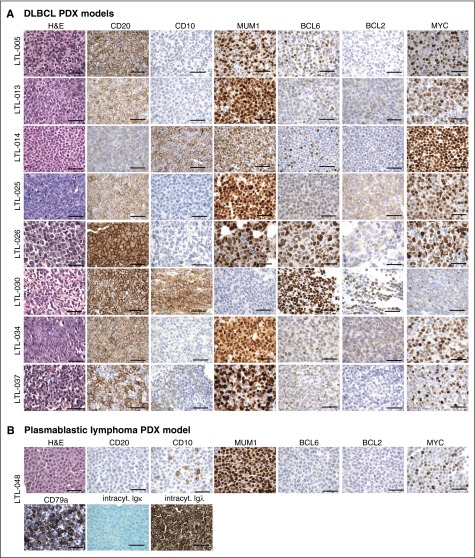
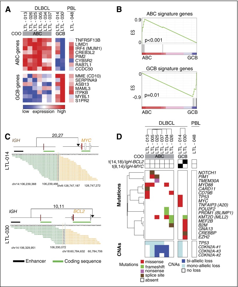
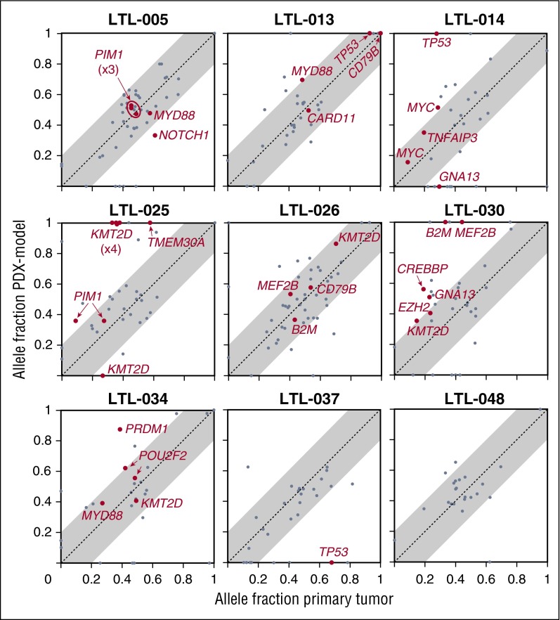
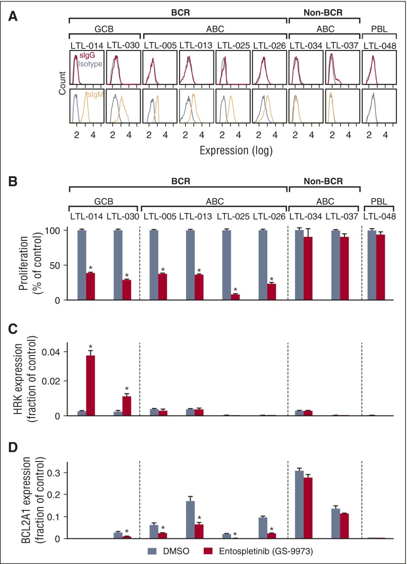
Diffuse large B-cell lymphoma (DLBCL) is a heterogeneous disease defined by transcriptional classifications, specific signaling and survival pathways, and multiple low-frequency genetic alterations. Preclinical model systems that capture the genetic and functional heterogeneity of DLBCL are urgently needed. Here, we generated and characterized a panel of large B-cell lymphoma (LBCL) patient-derived xenograft (PDX) models, including 8 that reflect the immunophenotypic, transcriptional, genetic, and functional heterogeneity of primary DLBCL and 1 that is a plasmablastic lymphoma. All LBCL PDX models were subjected to whole-transcriptome sequencing to classify cell of origin and consensus clustering classification (CCC) subtypes. Mutations and chromosomal rearrangements were evaluated by whole-exome sequencing with an extended bait set. Six of the 8 DLBCL models were activated B-cell (ABC)-type tumors that exhibited ABC-associated mutations such as MYD88, CD79B, CARD11, and PIM1. The remaining 2 DLBCL models were germinal B-cell type, with characteristic alterations of GNA13, CREBBP, and EZH2, and chromosomal translocations involving IgH and either BCL2 or MYC Only 25% of the DLBCL PDX models harbored inactivating TP53 mutations, whereas 75% exhibited copy number alterations of TP53 or its upstream modifier, CDKN2A, consistent with the reported incidence and type of p53 pathway alterations in primary DLBCL. By CCC criteria, 6 of 8 DLBCL PDX models were B-cell receptor (BCR)-type tumors that exhibited selective surface immunoglobulin expression and sensitivity to entospletinib, a recently developed spleen tyrosine kinase inhibitor. In summary, we have established and characterized faithful PDX models of DLBCL and demonstrated their usefulness in functional analyses of proximal BCR pathway inhibition.


Figures

Figure 1
Figure 1
Establishment of 9 LBCL PDX models. (A) Hematoxylin and eosin stains of 9 LBCL PDX models propagated under the renal capsule of NSG mice. Original magnification ×200. (B) Epstein-Barr-encoded RNA in situ hybridization of all 9 LBCL PDX models. An EBV-positive classical Hodgkin lymphoma (cHL) served as positive control. Bars represent 100 μm. (C) Clonality of the 9 PDX models assessed and interpreted according to the EuroClonality/BIOMED-2 guidelines. An IgH VH-JH spanning PCR (FR2, left) was performed on all 9 LBCL PDX models. Tumors without a clonal peak (LTL-005, LTL-037, and LTL-048) or an inconclusive result (LTL-037) were sequentially analyzed with a second PCR (FR1, right). Genomic DNA (gDNA) of NSG mouse tail served as a negative control, gDNA of peripheral blood mononuclear cells from a healthy human volunteer served as a polyclonal control, and gDNA of the Burkitt lymphoma cell line (BL30) served as a monoclonal control. EBV, Epstein-Barr virus; FR, fragment; PCR, polymerase chain reaction.
Figure 2
Figure 2
IHC characterization of all 9 PDX models. (A) IHC analyses of the indicated markers in all 8 DLBCL PDX models, which were consistent with the diagnosis of DLBCL. (B) IHC assessment of indicated markers in PDX model LTL-048, which is consistent with the diagnosis of PBL. Scale bars, 100 μm. See also Table 1. intracyt., intracytoplasmic.
Figure 3
Figure 3
Molecular heterogeneity of LBCL PDX models. (A) Heatmap of the relative expression of ABC- and GCB-signature genes in ABC- and GCB-type DLBCL PDX models and the additional PBL PDX model. Note that reads aligned to the mouse genome were filtered before the DLBCL PDX models were classified by COO. (B) Gene set enrichment analyses of the ABC-signature genes (upper) and GCB-signature genes (lower) in the ABC- and GCB-type DLBCL PDX models. (C) Detected IGH-MYC (upper) and IGH-BCL2 (lower) translocations in LTL-014 and LTL-030, respectively. Translocations are plotted in their genomic context. Exons are visualized as boxes, with ATG-containing exons in red, coding regions underlined in green, and enhancer regions underlined in black. Numbers of supporting reads (split reads, read pairs) are indicated above, and individual supporting reads are shown below. (D) Protein-perturbing mutations with an allele fraction >0.1 in the LBCL models. Mutations in most frequently reported recurrently mutated genes in primary DLBCL (supplemental Table 2C) are visualized as a color-coded heatmap (red, missense mutation; green, frameshift mutation; purple, nonsense mutation; brown, splice site mutation; white, mutation absent); CNAs in TP53 and CDKN2A are represented as a color-coded heatmap (dark blue, biallelic loss; light blue, monoallelic loss; white, no loss). COO transcriptional subtypes and identified translocations juxtaposing either BCL2 or MYC to the IgH locus are indicated in the legend above of the heatmap. ES, enrichment score.
Figure 4
Figure 4
Comparison of mutant allele fraction between primary tumor and associated PDX model. We plotted the mutant allele fraction (frequency of a mutation at a particular locus) in the primary tumors (x-axis) and the associated PDX models (y-axis). SNVs in genes reported to be mutated in at least 1 of 4 DLBCL sequencing series are represented in gray, and SNVs in genes reported to be mutated in at least 2 of 4 DLBCL sequencing series are labeled in red (supplemental Table 2A-). Multiple SNVs in the same gene are noted (PIM1 in LTL-005, KMT2D in LTL-025, and POU2F2 in LTL-034). Mutant allele fractions along the diagonal (x = y, identical mutant allele fractions, dotted line) indicate similar clonal frequencies in the primary tumor and associated PDX model. Mutant alleles that are more abundant in the PDX model than in the primary tumors are above the diagonal. Allele fractions ±0.2 from the identical mutant allele fractions are visualized in gray.
Figure 5
Figure 5
Analyses of cell surface immunoglobulin and BCR signaling in the LBCL PDX models. (A) Single-cell suspensions from each LBCL PDX model were gated for human CD45-positive cells and analyzed for surface immunoglobulin (IgG, red; IgM, orange; isotype, gray). CCC and COO subtypes of each model are indicated above the flow histograms. (B) Cellular proliferation of PDX tumor cell suspensions after chemical SYK inhibition with entospletinib (GS-9973) for 24 hours. (C,D) HRK (C) and BCL2A1 (D) transcript abundance following entospletinib (GS-9973) treatment. Experiments were performed in triplicate. A representative experiment of biological duplicates in shown. DMSO, dimethylsulfoxide. The P values were obtained using a Student t test (*P < .05).

Similar articles

  • Simplified algorithm for genetic subtyping in diffuse large B-cell lymphoma.
    Shen R, Fu D, Dong L, Zhang MC, Shi Q, Shi ZY, Cheng S, Wang L, Xu PP, Zhao WL. Shen R, et al. Signal Transduct Target Ther. 2023 Apr 10;8(1):145. doi: 10.1038/s41392-023-01358-y. Signal Transduct Target Ther. 2023. PMID: 37032379 Free PMC article.
  • Genetics and Pathogenesis of Diffuse Large B-Cell Lymphoma.
    Schmitz R, Wright GW, Huang DW, Johnson CA, Phelan JD, Wang JQ, Roulland S, Kasbekar M, Young RM, Shaffer AL, Hodson DJ, Xiao W, Yu X, Yang Y, Zhao H, Xu W, Liu X, Zhou B, Du W, Chan WC, Jaffe ES, Gascoyne RD, Connors JM, Campo E, Lopez-Guillermo A, Rosenwald A, Ott G, Delabie J, Rimsza LM, Tay Kuang Wei K, Zelenetz AD, Leonard JP, Bartlett NL, Tran B, Shetty J, Zhao Y, Soppet DR, Pittaluga S, Wilson WH, Staudt LM. Schmitz R, et al. N Engl J Med. 2018 Apr 12;378(15):1396-1407. doi: 10.1056/NEJMoa1801445. N Engl J Med. 2018. PMID: 29641966 Free PMC article.
  • Distinct molecular profile of IRF4-rearranged large B-cell lymphoma.
    Ramis-Zaldivar JE, Gonzalez-Farré B, Balagué O, Celis V, Nadeu F, Salmerón-Villalobos J, Andrés M, Martin-Guerrero I, Garrido-Pontnou M, Gaafar A, Suñol M, Bárcena C, Garcia-Bragado F, Andión M, Azorín D, Astigarraga I, Sagaseta de Ilurdoz M, Sábado C, Gallego S, Verdú-Amorós J, Fernandez-Delgado R, Perez V, Tapia G, Mozos A, Torrent M, Solano-Páez P, Rivas-Delgado A, Dlouhy I, Clot G, Enjuanes A, López-Guillermo A, Galera P, Oberley MJ, Maguire A, Ramsower C, Rimsza LM, Quintanilla-Martinez L, Jaffe ES, Campo E, Salaverria I. Ramis-Zaldivar JE, et al. Blood. 2020 Jan 23;135(4):274-286. doi: 10.1182/blood.2019002699. Blood. 2020. PMID: 31738823 Free PMC article.
  • Molecular Pathogenesis of Diffuse Large B-Cell Lymphoma.
    Dobashi A. Dobashi A. J Clin Exp Hematop. 2016;56(2):71-78. doi: 10.3960/jslrt.56.71. J Clin Exp Hematop. 2016. PMID: 27980305 Free PMC article. Review.
  • Rewired NFκB signaling as a potentially actionable feature of activated B-cell-like diffuse large B-cell lymphoma.
    Knittel G, Liedgens P, Korovkina D, Pallasch CP, Reinhardt HC. Knittel G, et al. Eur J Haematol. 2016 Dec;97(6):499-510. doi: 10.1111/ejh.12792. Epub 2016 Sep 8. Eur J Haematol. 2016. PMID: 27526684 Review.

Cited by

References

    1. Freedman AS, Friedberg JW, Aster JC. Classification of the hematopoietic neoplasms. Available at: www.uptodate.com/contents/classification-of-the-hematopoietic-neoplasms?.... Accessed August 9, 2015.
    1. Monti S, Savage KJ, Kutok JL, et al. Molecular profiling of diffuse large B-cell lymphoma identifies robust subtypes including one characterized by host inflammatory response. Blood. 2005;105(5):1851–1861. - PubMed
    1. Wright G, Tan B, Rosenwald A, Hurt EH, Wiestner A, Staudt LM. A gene expression-based method to diagnose clinically distinct subgroups of diffuse large B cell lymphoma. Proc Natl Acad Sci USA. 2003;100(17):9991–9996. - PMC - PubMed
    1. Caro P, Kishan AU, Norberg E, et al. Metabolic signatures uncover distinct targets in molecular subsets of diffuse large B cell lymphoma. Cancer Cell. 2012;22(4):547–560. - PMC - PubMed
    1. Chen L, Monti S, Juszczynski P, et al. SYK inhibition modulates distinct PI3K/AKT- dependent survival pathways and cholesterol biosynthesis in diffuse large B cell lymphomas. Cancer Cell. 2013;23(6):826–838. - PMC - PubMed

Publication types