. 2016 Feb 15;196(4):1855-64.
doi: 10.4049/jimmunol.1501802. Epub 2016 Jan 15.

Inducible Expression of CXCL1 within the Central Nervous System Amplifies Viral-Induced Demyelination

Affiliations

Inducible Expression of CXCL1 within the Central Nervous System Amplifies Viral-Induced Demyelination

Brett S Marro et al. J Immunol. .

Abstract

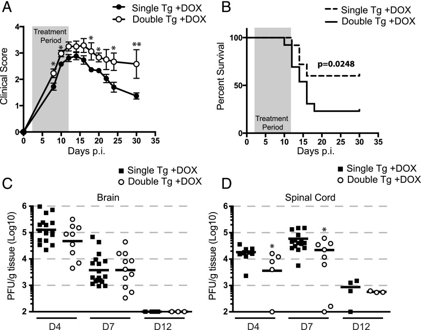
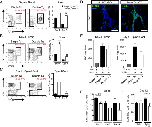
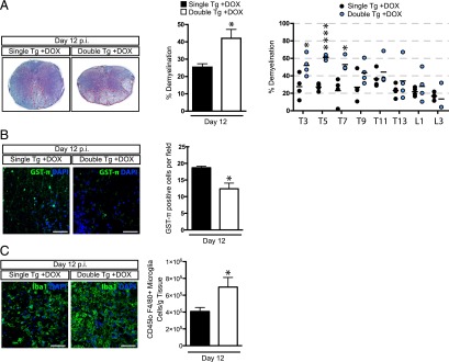
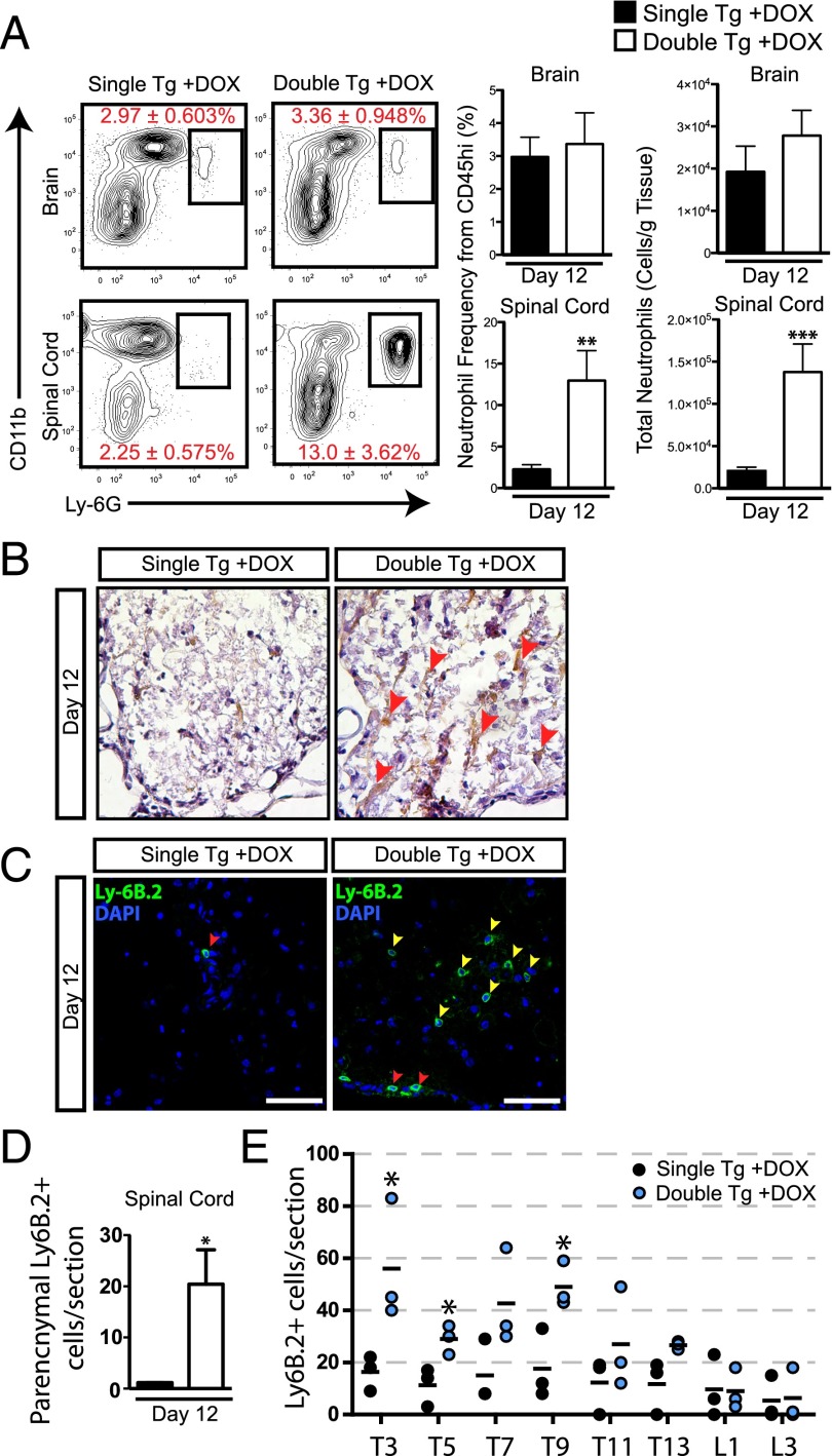
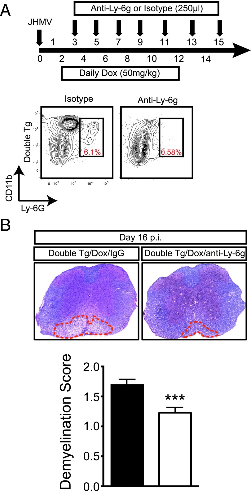
The functional role of the ELR(+) chemokine CXCL1 in host defense and disease following infection of the CNS with the neurotropic JHM strain of mouse hepatitis virus (JHMV) was examined. Mice in which expression of CXCL1 is under the control of a tetracycline-inducible promoter active within glial fibrillary acidic protein-positive cells were generated and this allowed for selectively increasing CNS expression of CXCL1 in response to JHMV infection and evaluating the effects on neuroinflammation, control of viral replication, and demyelination. Inducible expression of CNS-derived CXCL1 resulted in increased levels of CXCL1 protein within the serum, brain, and spinal cord that correlated with increased frequency of Ly6G(+)CD11b(+) neutrophils present within the CNS. Elevated levels of CXCL1 did not influence the generation of virus-specific T cells, and there was no difference in control of JHMV replication compared with control mice, indicating that T cell infiltration into the CNS is CXCL1-independent. Sustained CXCL1 expression within the CNS resulted in increased mortality that correlated with elevated neutrophil infiltration, diminished numbers of mature oligodendrocytes, and an increase in the severity of demyelination. Neutrophil ablation in CXCL1-transgenic mice reduced the severity of demyelination in mice, arguing for a role for these cells in white matter damage. Collectively, these findings illustrate that sustained CXCL1 expression amplifies the severity of white matter damage and that neutrophils can contribute to this process in a model of viral-induced neurologic disease.


Figures

FIGURE 1.
FIGURE 1.
Generation and in vitro characterization of a Dox-inducible, astrocyte-specific CXCL1 overexpressing mouse line. (A) Double-tg mice utilizing the tetracycline operon–controlled Dox-inducible system were generated by crossing hemizygous human GFAP-rtTA*M2 mice to a tg mouse line incorporating a tetracycline responsive element driving expression of CXCL1 (pBI-CXCL1). Shown are PCR results with genomic DNA from mice revealing amplicons specific for either pBI-CXCL1 or GFAP-rtTa. The internal control was performed when detecting GFAP-rtTa amplicons according to defined Jackson Laboratory protocols. (B) Cortex tissue from double-tg and single-tg postnatal day 1 mice was dissociated and enriched for astrocytes. Following 24 h of Dox (100 ng/ml)-treated double-tg astrocyte cultures, immunofluorescence (original magnification ×20) confirmed CXCL1 expression within GFAP+ astrocytes whereas vehicle treatment yielded no CXCL1 fluorescence. (C) Supernatants from Dox-treated double-tg and single-tg astrocytes cultures were collected and levels of CXCL1 were measured by ELISA. In the presence of Dox, double-tg cultures yielded significantly elevated CXCL1 protein levels, whereas single-tg cultures did not. Data from (C) represent supernatants pooled from triplicate wells derived from three separate double-tg and single-tg mouse cultures. Data are presented as average ± SEM; statistical significant was measured with an unpaired two-tailed Student t test. **p < 0.01.
FIGURE 2.
FIGURE 2.
CXCL1 is induced in vivo following administration of Dox into double-tg mice during acute JHMV infection. (A) To test the ability of Dox to induce CXCL1 overproduction in vivo, double-tg and single-tg mice were infected with 250 PFU JHMV and treated daily with 50 mg/kg Dox between days 2 and 12 p.i. (B) Administration of Dox to double-tg mice resulted in a significant increase in the expression of CXCL1 mRNA compared with Dox-treated single-tg mice within the brain at days 4, 7, and 12 p.i. as measured by quantitative real-time PCR. (C) Within the spinal cord, dox-treated double-tg mice had statistically significant increases in CXCL1 mRNA expression over Dox-treated single-tg mice at days 7 and 12 p.i. (D) CXCL1 transgene expression within the brain and spinal cord did not impact endogenous CXCL1 production within Dox-treated double-tg mice compared with Dox-treated single-tg mice. Overproduction of CXCL1 protein was observed within the spinal cord at day 7 p.i. within double-tg mice (E), and this correlated with an increase in CXCL1 protein within the blood serum (F) at day 7 p.i. as measured by ELISA. (G) Immunofluorscence analysis (original magnification ×40) of spinal cord tissue from Dox-treated double-tg mice confirmed that astrocytes were the source of CXCL1 production at day 7 p.i. (H) No significant changes in proinflammatory gene expression between double-tg and single-tg mice were observed within the spinal cord at day 12 p.i. using an RNA superarray. Data from (B) represent two experiments with a minimum of four mice per group. All quantitative real-time PCR samples were run in triplicate. (D) and (E) represent a minimum of three mice per group. Superarray data were compiled used the average value of two mice per group run in duplicate. Data are presented as average ± SEM; statistical significance was measured using an unpaired two-tailed Student t test. *p < 0.05, **p < 0.01.
FIGURE 3.
FIGURE 3.
CXCL1 overproduction amplifies JHMV-induced clinical disease and mortality but is not a result of delayed viral clearance. Double-tg and single-tg mice were infected with 250 PFU JHMV and treated with 50 mg/kg Dox daily starting at day 2 p.i. through day 12 p.i. (A) Clinical severity was assessed until day 30 p.i. using a four-point scale. (B) Dox treatment of double-tg mice led to significantly increased mortality compared with Dox-treated single-tg controls. Viral titers of Dox-treated double-tg and single-tg mice were measured within both the brain (C) and spinal cord (D) by plaque assay. Each symbol represents one individual mouse; solid black lines represent geometric means. Statistical significance was measured with the Mann–Whitney U test. Clinical disease and mortality scoring represents an average of double-tg and single-tg mice. For (A) and (B), n = 23 single-tg mice and n = 12 double-tg mice. Statistical significance for the clinical scoring data was determined by a Student t test for each time point assessed. Statistical comparison of the survival curves was measured using the Mantel–Cox test. *p < 0.05, **p < 0.01.
FIGURE 4.
FIGURE 4.
CXCL1 overproduction from astrocytes mobilizes neutrophils and directs them to the CNS. Blood from Dox-treated, JHMV-infected double-tg and single-tg mice was isolated at day 4 and day 7 p.i. and used for flow cytometric analysis. (A) The frequency of CD11b+Ly6G+ neutrophils was significantly higher within the blood at days 4 and 7 p.i. Neutrophil migration to the brains (B) and spinal cords (C) of both groups was assessed by flow cytometry at days 4 and 7 p.i. A significant increase in neutrophil frequency within the brain and spinal cord was observed in Dox-treated double-tg mice at both time points. (D) Immunofluorescence analysis indicated that Ly6B.2+ neutrophils were primarily located at the ependymal lining and perivascular space at the spinal cord, with minimal neutrophil presence within the parenchyma. Scale bars, 50 μM. (E) At day 4 p.i., brains and spinal cords from Dox-treated single-tg and double-tg mice treated with NaFl were removed and total NaFl uptake was measured. No differences in the frequency of CD11b+Ly6C+Ly6G cells were detected within either the blood (F) or brain and spinal cords (G) between Dox-treated double-tg or single-tg mice at defined times after infection with JHMV. (A)–(C) represent three independent experiments with a minimum of three mice per group per experiment at each time point analyzed; (F) and (G) represent two independent experiments with a minimum of three mice per group per experiment at each time point analyzed. Data are presented as average ± SEM; statistical significance was measured using an unpaired two-tailed Student t test. *p < 0.05, **p < 0.01, ***p < 0.001, ****p < 0.0001.
FIGURE 5.
FIGURE 5.
Elevated CXCL1 expression is associated with increased demyelination. Histopathological analysis of spinal cords of double-tg mice reveals an increase in demyelination. (A) Representative Luxol fast blue–stained spinal cords (original magnification ×4) reveal increased (p < 0.05) demyelination in Dox-treated double-tg mice compared with single-tg controls. Assessment of demyelination in defined spinal cord sections indicates white matter damage is more concentrated within the upper thoracic (T) regions (T3–T7) compared with lower T9–T13 as well as lumbar (L) regions examined. (B) Increased demyelination was associated with a reduction in the number total GST-π+ oligodendrocytes within the white matter. The dot plot presented in (A) represents the percentage demyelination at specific anatomical regions of the spinal cord. (C) Immunofluorescence staining for Iba1 revealed increased numbers of positive cells within areas of demyelination in spinal cords of double-tg compared with single-tg mice at day 12 p.i., and flow analysis indicated increased (p < 0.05) numbers of microglia (CD45loF4/80+). Scale bars, 50 μM. (A)–(C) represent a minimum of four mice per group. Data are presented as average ± SEM; statistical significance was measured using an unpaired two-tailed Student t test. *p < 0.05.
FIGURE 6.
FIGURE 6.
Neutrophils are found within parenchymal regions of the spinal cord in double-tg mice. (A) At day 12 p.i. brains and spinal cords were isolated and single-cell suspensions were prepared from double-tg and single-tg mice infected with JHMV. Flow cytometric analysis revealed a significant increase in the frequency and total number of neutrophils within the spinal cord of double-tg mice. (B) CXCL1 protein was detected by immunohistochemical staining (red arrowheads) within spinal cords of Dox-treated double-tg mice, and (C) immunofluorescence staining further demonstrated a significant increase in the number of Ly6B.2+ neutrophils (yellow arrowheads). Scale bars, 50 μM. (D) Quantification of neutrophils within the spinal cords indicated an overall increase (p < 0.05) in Dox-treated double-tg mice compared with Dox-treated single-tg mice. (E) Quantification of neutrophils in defined anatomic regions of spinal cords showed increased numbers of neutrophils (p < 0.05) within upper thoracic (T) regions (T3–T9) and lower numbers at T11–T13 and lumbar (L) regions in Dox-treated double-tg mice compared with Dox-treated single-tg mice. (A) represents three independent experiments with a minimum of three mice per group per experiment. (D) and (E) represent a minimum of three mice per group. Data are presented as average ± SEM; statistical significance was measured using an unpaired two-tailed Student t test. *p < 0.05.
FIGURE 7.
FIGURE 7.
Neutrophils amplify the severity of demyelination. (A) Dox-treated JHMV-infected double-tg mice were treated with either anti-Ly6g Ab or isotype-matched control starting between days 3 and 4 p.i. and continuing every other day until day 15 p.i. Representative flow analysis of spinal cords confirmed anti-Ly6G treatment successfully depleted neutrophils. (B) Representative Luxol fast blue–stained spinal cord sections from JHMV-infected double-tg mice treated with either control IgG2a or anti-Ly6G Ab between days 3 and 15 p.i. Quantification of the severity of demyelination revealed reduced white matter damage in mice treated with anti-Ly6G Ab compared with mice treated with isogenic IgG2a control Ab; data are derived from two independent experiments with a minimum of three mice per group per experiment. Data are presented as average ± SEM; statistical significance was measured using an unpaired two-tailed Student t test. *p < 0.001.

Similar articles

Cited by

References

    1. Bergmann C. C., Lane T. E., Stohlman S. A. 2006. Coronavirus infection of the central nervous system: host-virus stand-off. Nat. Rev. Microbiol. 4: 121–132. - PMC - PubMed
    1. Liu M. T., Chen B. P., Oertel P., Buchmeier M. J., Armstrong D., Hamilton T. A., Lane T. E. 2000. The T cell chemoattractant IFN-inducible protein 10 is essential in host defense against viral-induced neurologic disease. J. Immunol. 165: 2327–2330. - PubMed
    1. Lane T. E., Liu M. T., Chen B. P., Asensio V. C., Samawi R. M., Paoletti A. D., Campbell I. L., Kunkel S. L., Fox H. S., Buchmeier M. J. 2000. A central role for CD4+ T cells and RANTES in virus-induced central nervous system inflammation and demyelination. J. Virol. 74: 1415–1424. - PMC - PubMed
    1. Hosking M. P., Liu L., Ransohoff R. M., Lane T. E. 2009. A protective role for ELR+ chemokines during acute viral encephalomyelitis. PLoS Pathog. 5: e1000648. - PMC - PubMed
    1. Lane T. E., Asensio V. C., Yu N., Paoletti A. D., Campbell I. L., Buchmeier M. J. 1998. Dynamic regulation of α- and β-chemokine expression in the central nervous system during mouse hepatitis virus-induced demyelinating disease. J. Immunol. 160: 970–978. - PubMed

Publication types

MeSH terms