Review
doi: 10.17305/bjbms.2016.917.
The mouse prostate: a basic anatomical and histological guideline
Affiliations
- PMID: 26773172
- PMCID: PMC4765945
- DOI: 10.17305/bjbms.2016.917
Item in Clipboard
Review
The mouse prostate: a basic anatomical and histological guideline
Bosn J Basic Med Sci.
.
Abstract
Despite substantial similarities in embryological, cellular and molecular biology features, human and mouse prostates differ in organ morphology and tissue architecture. Thus, a clear understanding of the anatomy and histology of the mouse prostate is essential for the identification of urogenital phenotypes in genetically engineered mice, as well as for the study of the etiology, development, and treatment of human prostatic diseases for which mouse models are used. The purpose of this manuscript is to provide a brief guide for the dissection of the mouse prostate and the identification of its different lobes and histology, to both basic researchers and medical pathologists who are unfamiliar with mouse tissues.
Figures
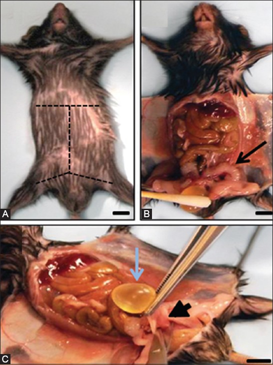
Mouse dissection and excision of the male urogenital tract. (A) Adult male mouse in supine position. Dashed lines indicate the skin incision pattern to follow; (B) Bladder (below cotton swab) and seminal vesicles (arrow) are easily identified once the abdominal cavity of the mouse is opened; (C) Most of the urogenital tract can be harvested from the mouse carcass en bloc by grasping the bladder with tweezers, pulling it up, and cutting the base of the urethra (arrowhead). Scale bar, 0.5 cm.
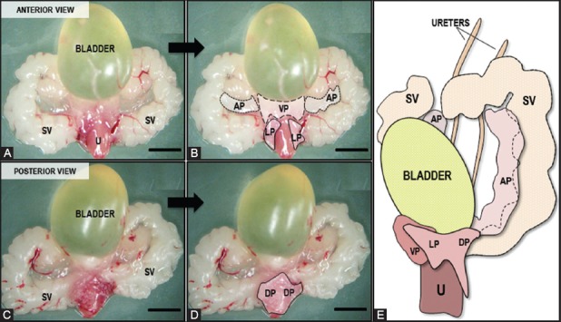
Gross view of mouse male urogenital organs. (A) Anterior and (C) posterior aspects of the urogenital tract dissected from an adult male mouse, as seen under the dissecting microscope. Urethra (U), Seminal vesicles (SV); (B) and (D) show the same images with dashed and dotted lines delineating the ventral (VP), lateral (LP), anterior (AP), and dorsal (DP) prostate lobes; (E) Anterolateral view (left side) of the prostate lobes relative to the bladder, seminal vesicles, and urethra (Modified from: Sugimura, Y., et al. [15]). Scale bar, 1 cm.
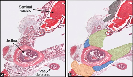
Cross section of a normal urogenital tract from an adult male mouse. (A) Adjacent to the prostate lobes here can be seen the urethra and a vas deferens, both surrounded by thick muscular walls, and a seminal vesicle with characteristic large lumen filled with strong esosinophilic secretion; (B) The different prostate lobes have been highlighted in orange (VP), light blue (LP), green (DP), and grey (AP), for proper identification. Hematoxylin and eosin, 25X.
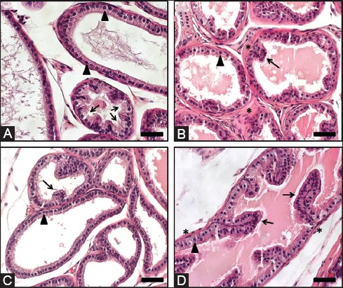
Mouse prostate lobes. Representative images are shown. (A) Ventral prostate: moderate to large acini are lined by cuboidal to simple columnar epithelium with flat luminal borders. Note the basally located nuclei (arrowheads) and the tufting pattern (arrows). A pale serous secretion is a unique characteristic of the ventral prostate lobe; (B) Dorsal Prostate: small acini are mostly line by simple columnar epithelium with centrally located nuclei (arrowhead) and sparse infoldings (arrow). A dense stroma (*) surrounding the acini is typical. An eosinophilic secretion is typically seen in the acini; (C) Lateral prostate: small to large acini are lined by cuboidal to low columnar epithelium with nuclei located basally (arrowhead) that form sparse infoldings (arrow). Similar to the ventral prostate, acini from the lateral prostate have a flat luminal border; (D) Anterior prostate: moderate to large acini lined mostly by cuboidal to columnar epithelium with nuclei located centrally (arrowhead) and with infoldings (arrows). Abundant eosinophilic luminal secretion is characteristic, along with the presence of noticeable smooth muscle (*) surrounding each acinus. Hematoxylin and eosin. Scale bar, 100 µm.

Identification of mouse prostate lobes. This decision tree suggests some key points that the basic researcher or pathologist may use to identify the different mouse prostate lobes in hematoxylin and eosin stained sections of the male mouse genitourinary tract.
Similar articles
-
Ventral prostate fibrosis in the Akita mouse is associated with macrophage and fibrocyte infiltration.J Diabetes Res. 2014;2014:939053. doi: 10.1155/2014/939053. Epub 2014 Jun 11. J Diabetes Res. 2014. PMID: 25019092 Free PMC article.
-
Overview of prostate anatomy, histology, and pathology.Endocrinol Metab Clin North Am. 2011 Sep;40(3):565-75, viii-ix. doi: 10.1016/j.ecl.2011.05.012. Endocrinol Metab Clin North Am. 2011. PMID: 21889721 Review.
-
Genetically engineered mouse models to study prostate cancer.Methods Mol Biol. 2015;1267:73-91. doi: 10.1007/978-1-4939-2297-0_4. Methods Mol Biol. 2015. PMID: 25636465
-
Identification, Histological Characterization, and Dissection of Mouse Prostate Lobes for In Vitro 3D Spheroid Culture Models.J Vis Exp. 2018 Sep 18;(139):58397. doi: 10.3791/58397. J Vis Exp. 2018. PMID: 30295668 Free PMC article.
-
Anatomy and Histology of the Human and Murine Prostate.Cold Spring Harb Perspect Med. 2018 May 1;8(5):a030346. doi: 10.1101/cshperspect.a030346. Cold Spring Harb Perspect Med. 2018. PMID: 29038334 Free PMC article. Review.
Cited by
-
Impact of sex, androgens, and prostate size on C57BL/6J mouse urinary physiology: urethral histology.Am J Physiol Renal Physiol. 2020 Mar 1;318(3):F617-F627. doi: 10.1152/ajprenal.00540.2019. Epub 2020 Jan 6. Am J Physiol Renal Physiol. 2020. PMID: 31904290 Free PMC article.
-
Impact of sex, androgens, and prostate size on C57BL/6J mouse urinary physiology: functional assessment.Am J Physiol Renal Physiol. 2019 Oct 1;317(4):F996-F1009. doi: 10.1152/ajprenal.00270.2019. Epub 2019 Aug 7. Am J Physiol Renal Physiol. 2019. PMID: 31390231 Free PMC article.
-
White button mushroom (Agaricus bisporus) disrupts androgen receptor signaling in human prostate cancer cells and patient-derived xenograft.J Nutr Biochem. 2021 Mar;89:108580. doi: 10.1016/j.jnutbio.2020.108580. Epub 2020 Dec 31. J Nutr Biochem. 2021. PMID: 33388344 Free PMC article.
-
Structural and Ultrastructural Morphological Evaluation of Giant Anteater (Myrmecophaga tridactyla) Prostate Gland.Biology (Basel). 2021 Mar 17;10(3):231. doi: 10.3390/biology10030231. Biology (Basel). 2021. PMID: 33802717 Free PMC article.
-
Morphologic description of male reproductive accessory glands in a mouse model of mucopolysaccharidosis type I (MPS I).J Mol Histol. 2020 Apr;51(2):137-145. doi: 10.1007/s10735-020-09864-x. Epub 2020 Mar 11. J Mol Histol. 2020. PMID: 32162173
References
-
- Roehrborn C. Insights into the relationships between prostatic disorders and their potential impact on future urologic practice. Eur Urol. 2013;5(Suppl):698–703.
-
- Luke MC, Coffey DS. The male sex accessory tissues. In: Knobil E, Nelly JD, editors. The Physiology of Reproduction. New York: Raven Press; 1994. pp. 1435–89.
-
- Marker PC, Donjacour AA, Dahiya R, Cunha GR. Hormonal, cellular, and molecular control of prostatic development. Dev Biol. 2003;253(2):165–74. http://dx.doi.org/10.1016/S0012-1606(02)00031-3 . - PubMed
-
- Timms BG, Mohs TJ, Didio LJ. Ductal budding and branching patterns in the developing prostate. J Urol. 1994;151(5):1427–32. - PubMed
-
- Prins GS, Lindgren M. Accessory sex glands in the male. In: Plant TM, Zeleznik AJ, editors. Knobil and Neill’s physiology of reproduction. 14th ed. London, UK: Elsevier; 2015. pp. 773–804. http://dx.doi.org/10.1016/b978-0-12-397175-3.00018-1 .
Publication types
MeSH terms
Substances
LinkOut - more resources
Full Text Sources
Other Literature Sources