The role of Six1 signaling in paclitaxel-dependent apoptosis in MCF-7 cell line
- PMID: 26773176
- PMCID: PMC4765936
- DOI: 10.17305/bjbms.2016.674
The role of Six1 signaling in paclitaxel-dependent apoptosis in MCF-7 cell line
Abstract
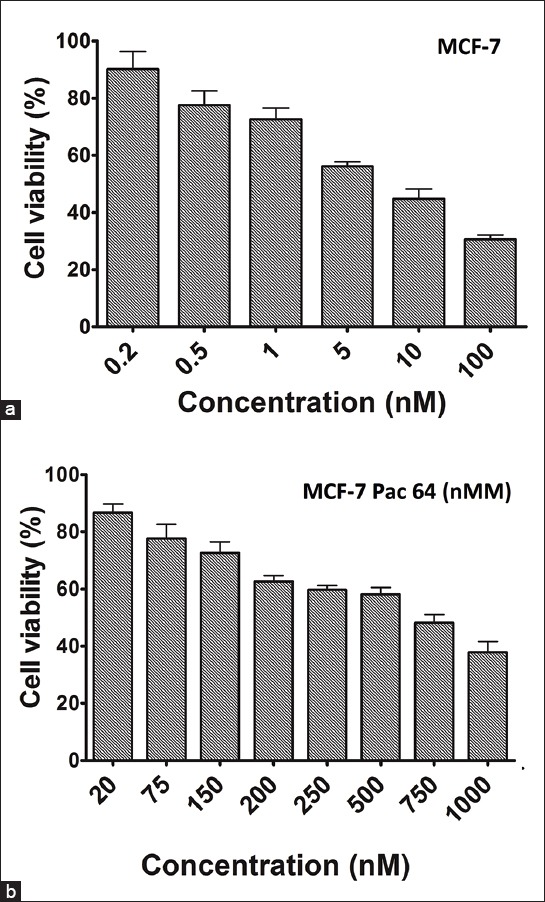
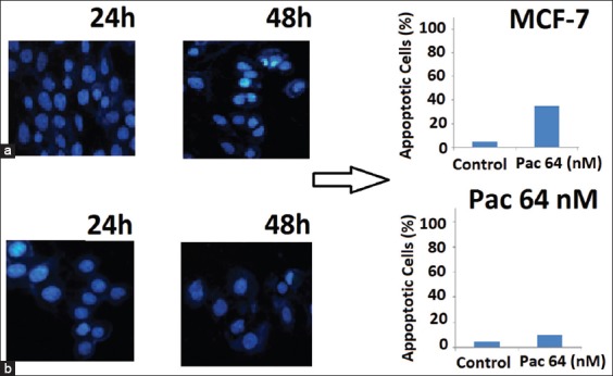
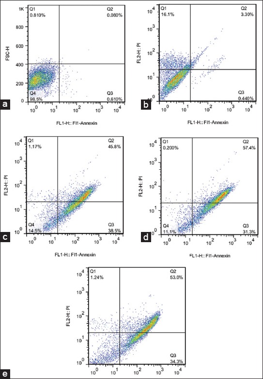
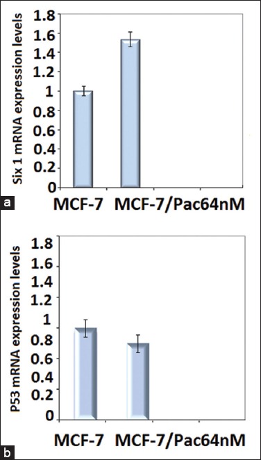
The resistance of cancer cells to chemotherapeutic agents represents the main problem in cancer treatment. Despite intensive research, mechanisms of resistance have not yet been fully elucidated. Six1 signaling has an important role in the expansion of progenitor cell populations during early embryogenesis. Six1 gene overexpression has been strongly associated with aggressiveness, invasiveness, and poor prognosis of different cancers. In this study, we investigated the role of Six1 signaling in resistance of MCF-7 breast cancer cells to taxanes. We first established in vitro paclitaxel-resistant MCF-7 breast cancer cells. Morphological modifications in paclitaxel-resistant cells were examined via light microscopic images and fluorescence-activated cell sorting analysis. Applying quantitative real-time polymerase chain reaction, we measured Six1, B-cell lymphoma/leukemia(BCL-2), BAX, and P53 mRNA expression levels in both non-resistant and resistant cells. Resistant cells were developed from the parent MCF-7 cells by applying increasing concentrations of paclitaxel up to 64 nM. The inhibitory concentration 50% value in resistant cells increased from 3.5 ± 0.03 to 511 ± 10.22 nM (p = 0.015). In paclitaxel-resistant cells, there was a significant increase in Six1 and BCL-2 mRNA levels (p = 0.0007) with a marked decrease in pro-apoptotic Bax mRNA expression level (p = 0.03); however, there was no significant change in P53 expression (p = 0.025). Our results suggest that identifying cancer patients with high Six1 expression and then inhibition of Six1 signaling can improve the efficiency of chemotherapeutic agents in the induction of apoptosis.
Figures
References
-
- Alphandéry E. Perspectives of breast cancer thermotherapies. J Cancer. 2014;5(6):472–9. http://dx.doi.org/10.7150/jca.8693 . - PMC - PubMed
-
- Tinoco G, Warsch S, Glück S, Avancha K, Montero AJ. Treating breast cancer in the 21st century:emerging biological therapies. J Cancer. 2013;4(2):117–32. http://dx.doi.org/10.7150/jca.4925 . - PMC - PubMed
-
- Tabasinezhad M, Samadi N, Ghanbari P, Mohseni M, Saei AA, Sharifi S, et al. Sphingosin 1-phosphate contributes in tumor progression. J Cancer Res Ther. 2013;9(4):556–63. http://dx.doi.org/10.4103/0973-1482.126446 . - PubMed
-
- Jordan MA, Wilson L. Microtubules as a target for anticancer drugs. Nat Rev Cancer. 2004;4(4):253–65. http://dx.doi.org/10.1038/nrc1317 . - PubMed
-
- Coley HM. Development of drug-resistant models. Methods Mol Med. 2004;88:267–73. - PubMed
Publication types
MeSH terms
Substances
LinkOut - more resources
Full Text Sources
Other Literature Sources
Medical
Research Materials
Miscellaneous