. 2016 Mar;186(3):511-6.
doi: 10.1016/j.ajpath.2015.11.005. Epub 2016 Jan 7.

Pre-Existing Mature Oligodendrocytes Do Not Contribute to Remyelination following Toxin-Induced Spinal Cord Demyelination

Affiliations

Pre-Existing Mature Oligodendrocytes Do Not Contribute to Remyelination following Toxin-Induced Spinal Cord Demyelination

Abbe H Crawford et al. Am J Pathol. 2016 Mar.

Abstract

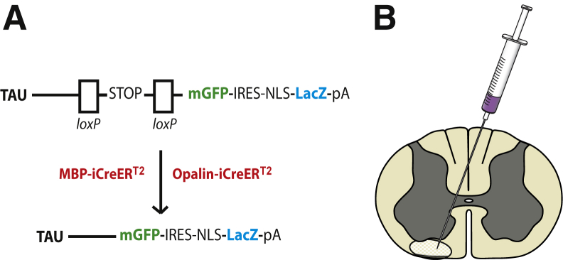
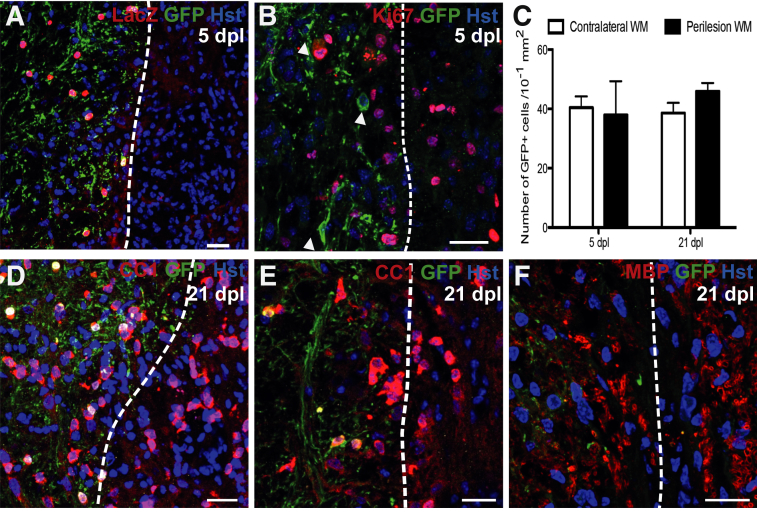
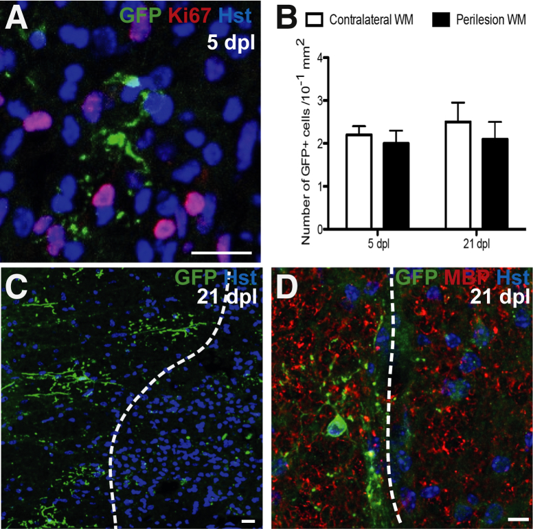
Remyelination is the regenerative response to demyelination. Although the oligodendrocyte progenitor is established as the major source of remyelinating cells, there is no conclusive evidence on whether mature, differentiated oligodendrocytes can also contribute to remyelination. Using two different inducible myelin-CreER mouse strains in which mature oligodendrocytes were prelabeled by the expression of membrane-bound Green fluorescent protein, we found that after focal spinal cord demyelination, the surrounding surviving labeled oligodendrocytes did not proliferate but remained at a consistent density. Furthermore, existing (prelabeled) oligodendrocytes showed no evidence of incorporation or migration into the lesioned area, or of process extension from the peripheral margins into the lesion. Thus, mature oligodendrocytes do not normally contribute to remyelination and are therefore not a promising target for regenerative therapy.


Figures

Figure 1
Figure 1
A: Transgenic mouse strains: A reporter mouse strain, in which membrane-bound Green fluorescent protein (mGfp) and β-galactosidase (LacZ) are under the control of the Tau promoter, was crossed to either of two inducible Cre strains in which Cre recombinase expression was restricted to mature oligodendrocytes using the Mbp or Opalin promoters. Excision of the STOP sequence by tamoxifen-inducible Cre recombination results in expression of mGfp and LacZ. B: Schematic diagram of the lysolecithin microinjection site in the white matter of the spinal cord ventral funiculus. IRES, internal ribosome entry site; NLS, nuclear localisation sequence; pA, polyadenylation.
Figure 2
Figure 2
A: Five days postlesion (dpl) in Mbp-iCreERT2:Tau-mGfp-LacZ mouse: dashed line indicates presumed lesion edge (based on increased cell density), with lesioned tissue on the right. B: Ki-67+ cells in the lesion and in perilesion white matter (WM) at 5 dpl: no surrounding Green fluorescent protein (Gfp)-positive oligodendrocytes (OLs) (indicated by arrowheads) costain for Ki-67. C: Number of Gfp+ cells in contralateral and perilesion WM at 5 and 21 dpl: no significant difference was detected. D–F: At 21 dpl, membranous Gfp+ processes do not extend into adjacent areas of remyelination. Adenomatous polyposis coli antibody hybridoma clone CC1–positive OLs: with images from two separate animals (D and E), and myelin basic protein (Mbp)-positive myelin sheaths (F) confirm remyelination of lesioned area. Data are expressed as means ± SEM. P = 0.69 at 5 dpl, P = 0.13 at 21 dpl (Student's t-test). Scale bars = 20 μm. Original magnification: ×20 (A); ×40 (B, D, and E); ×63 (F). Hst, hoechst.
Figure 3
Figure 3
A: Five days postlesion (dpl) in Opalin-iCreERT2:Tau-mGfp-LacZ mouse: Ki-67–Green fluorescent protein (Gfp)-positive oligodendrocyte (OL) from the presumed lesion edge surrounded by Ki-67+ cells. B: Number of membranous Gfp+cells in contralateral and perilesion white matter (WM) at 5 and 21 dpl: no significant difference is detected. C: Gfp+ OLs surrounding the lesion make no contribution to remyelination at 21 dpl (lesion right of dashed line). Fragments of Gfp+ staining are present within the lesion, but the morphology is not consistent with a myelinating cell and are presumed to be labeled myelin debris generated during demyelination that has not been cleared from the lesion. D: Gfp+ OL adjacent to remyelinated lesion: remyeination shown by myelin basic protein (Mbp)-positive myelin sheaths. Data are expressed as means ± SEM. P = 0.56 at 5 dpl, P = 0.75 at 21 dpl (t-test). Scale bars = 20 μm. Original magnification: ×63 (A and C); ×40 (B). Hst, hoechst.

References

    1. Zawadzka M., Rivers L.E., Fancy S.P., Zhao C., Tripathi R., Jamen F., Young K., Goncharevich A., Pohl H., Rizzi M., Rowitch D.H., Kessaris N., Suter U., Richardson W.D., Franklin R.J.M. CNS-resident glial progenitor/stem cells produce Schwann cells as well as oligodendrocytes during repair of CNS demyelination. Cell Stem Cell. 2010;6:578–590. - PMC - PubMed
    1. Tripathi R.B., Rivers L.E., Young K.M., Jamen F., Richardson W.D. NG2 glia generate new oligodendrocytes but few astrocytes in a murine experimental autoimmune encephalomyelitis model of demyelinating disease. J Neurosci. 2010;30:16383–16390. - PMC - PubMed
    1. Ludwin S.K. Proliferation of mature oligodendrocytes after trauma to the central nervous system. Nature. 1984;308:274–275. - PubMed
    1. Ludwin S.K., Bakker D.A. Can oligodendrocytes attached to nuclei proliferate? J Neurosci. 1988;8:1239–1244. - PMC - PubMed
    1. Wood P.M., Bunge R.P. Evidence that axons are mitogenic for oligodendrocytes isolated from adult animals. Nature. 1986;320:756–758. - PubMed

Publication types

LinkOut - more resources