Effect of growth hormone treatment on diastolic function in patients who have developed growth hormone deficiency after definitive treatment of acromegaly
- PMID: 26774401
- PMCID: PMC4716556
- DOI: 10.1016/j.ghir.2015.12.003
Effect of growth hormone treatment on diastolic function in patients who have developed growth hormone deficiency after definitive treatment of acromegaly
Abstract
Objective: Although growth hormone (GH) replacement is prescribed for patients with hypopituitarism due to many etiologies, it is not routinely prescribed for patients with GH deficiency (GHD) after cure of acromegaly (acroGHD). This study was designed to investigate the effect of GH replacement on cardiac parameters in acroGHD.
Design: We prospectively evaluated for 12months 23 patients with acroGHD: 15 subjects on GH replacement and eight subjects not on GH replacement. Main outcome measures included LV mass corrected for body surface area (LVM/BSA) and measures of diastolic dysfunction (E/A ratio and deceleration time), as assessed by echocardiography.
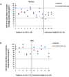
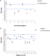
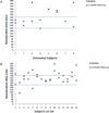
Results: After 12months of follow-up, there were no differences between the GH-treated group and the untreated group in LVM/BSA (GH: 74.4±22.5g/m(2) vs untreated: 72.9±21.3g/m(2), p=0.89), E/A ratio (GH: 1.21±0.39 vs untreated: 1.08±0.39, p=0.50) or deceleration time (GH: 224.5±60.1ms vs untreated: 260±79.8ms, p=0.32). The overall degree of diastolic function was similar between the groups with 42.9% of untreated subjects and 50% of GH-treated subjects (p=0.76) classified as having normal diastolic function at follow-up.
Conclusions: There were no significant differences in LVM/BSA or parameters of diastolic function in patients with a history of acromegaly treated for GHD as compared to those who were untreated. These data are reassuring with respect to cardiovascular safety with GH use after treatment for acromegaly, although further longer term study is necessary to evaluate the safety and efficacy of GH treatment in this population.
Keywords: Acromegaly; Cardiovascular risk; Diastolic dysfunction; Growth hormone deficiency.
Copyright © 2015 Elsevier Ltd. All rights reserved.
Conflict of interest statement
The authors declare that they have no conflict of interest.
Figures


Similar articles
-
Effects of long-term growth hormone replacement in adults with growth hormone deficiency following cure of acromegaly: a KIMS analysis.J Clin Endocrinol Metab. 2014 Jun;99(6):2018-29. doi: 10.1210/jc.2014-1013. Epub 2014 Apr 2. J Clin Endocrinol Metab. 2014. PMID: 24694339
-
Fractures in pituitary adenoma patients from the Dutch National Registry of Growth Hormone Treatment in Adults.Pituitary. 2016 Aug;19(4):381-90. doi: 10.1007/s11102-016-0716-3. Pituitary. 2016. PMID: 27048484 Free PMC article.
-
Effect of growth hormone replacement therapy on the quality of life in women with growth hormone deficiency who have a history of acromegaly versus other disorders.Endocr Pract. 2012 Mar-Apr;18(2):209-18. doi: 10.4158/EP11134.OR. Endocr Pract. 2012. PMID: 22440981 Free PMC article. Clinical Trial.
-
Growth hormone deficiency in treated acromegaly.Trends Endocrinol Metab. 2015 Jan;26(1):11-21. doi: 10.1016/j.tem.2014.10.005. Epub 2014 Nov 27. Trends Endocrinol Metab. 2015. PMID: 25434492 Review.
-
Growth hormone deficiency in treated acromegaly and active Cushing's syndrome.Best Pract Res Clin Endocrinol Metab. 2017 Feb;31(1):79-90. doi: 10.1016/j.beem.2017.03.002. Epub 2017 Mar 9. Best Pract Res Clin Endocrinol Metab. 2017. PMID: 28477735 Review.
Cited by
-
Filling the gap between the heart and the body in acromegaly: a case-control study.Endocrine. 2023 Feb;79(2):365-375. doi: 10.1007/s12020-022-03232-3. Epub 2022 Oct 30. Endocrine. 2023. PMID: 36309947 Free PMC article.
-
Heart in Acromegaly: The Echocardiographic Characteristics of Patients Diagnosed with Acromegaly in Various Stages of the Disease.Int J Endocrinol. 2018 Jul 11;2018:6935054. doi: 10.1155/2018/6935054. eCollection 2018. Int J Endocrinol. 2018. PMID: 30123265 Free PMC article.
References
-
- Rajasoorya C, Holdaway IM, Wrightson P, Scott DJ, Ibbertson HK. Determinants of clinical outcome and survival in acromegaly. Clin Endocrinol (Oxf) 1994;41:95–102. - PubMed
-
- Rosen T, Bengtsson BA. Premature mortality due to cardiovascular disease in hypopituitarism. Lancet. 1990;336:285–288. - PubMed
-
- Lopez-Velasco R, Escobar-Morreale HF, Vega B, Villa E, Sancho JM, Moya-Mur JL, Garcia-Robles R. Cardiac involvement in acromegaly: specific myocardiopathy or consequence of systemic hypertension? J Clin Endocrinol Metab. 1997;82:1047–1053. - PubMed
-
- Vilar L, Naves LA, Costa SS, Abdalla LF, Coelho CE, Casulari LA. Increase of classic and nonclassic cardiovascular risk factors in patients with acromegaly. Endocr Pract. 2007;13:363–372. - PubMed
-
- Hansen I, Tsalikian E, Beaufrere B, Gerich J, Haymond M, Rizza R. Insulin resistance in acromegaly: defects in both hepatic and extrahepatic insulin action. Am J Physiol. 1986;250:E269–E273. - PubMed
Publication types
MeSH terms
Substances
Grants and funding
LinkOut - more resources
Full Text Sources
Other Literature Sources
Medical

