. 2016 Jan 26;14(3):621-631.
doi: 10.1016/j.celrep.2015.12.045. Epub 2016 Jan 7.

Identifying Family-Member-Specific Targets of Mono-ARTDs by Using a Chemical Genetics Approach

Affiliations

Identifying Family-Member-Specific Targets of Mono-ARTDs by Using a Chemical Genetics Approach

Ian Carter-O'Connell et al. Cell Rep. .

Abstract

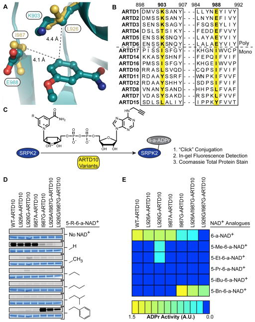
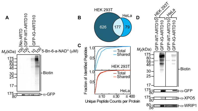
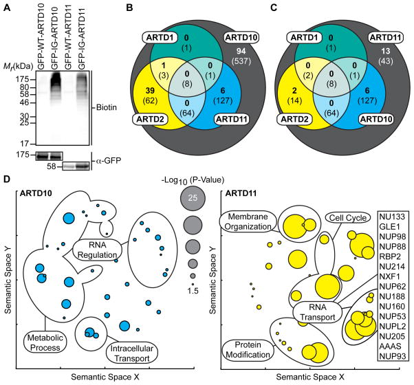
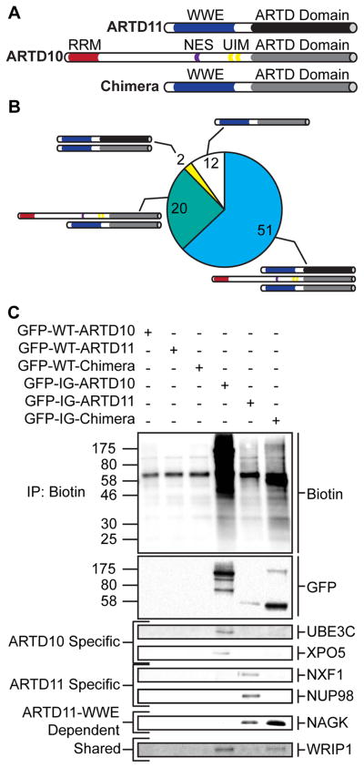
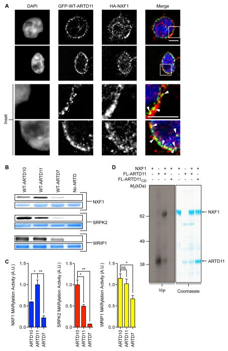
ADP-ribosyltransferases (ARTD1-16) have emerged as major downstream effectors of NAD(+) signaling in the cell. Most ARTDs (ARTD7 and 8, 10-12, and 14-17) catalyze the transfer of a single unit of ADP-ribose from NAD(+) to target proteins, a process known as mono-ADP-ribosylation (MARylation). Progress in understanding the cellular functions of MARylation has been limited by the inability to identify the direct targets for individual mono-ARTDs. Here, we engineered mono-ARTDs to use an NAD(+) analog that is orthogonal to wild-type ARTDs. We profiled the MARylomes of ARTD10 and ARTD11 in vitro, identifying isoform-specific targets and revealing a potential role for ARTD11 in nuclear pore complex biology. We found that ARTD11 targeting is dependent on both its regulatory and catalytic domains, which has important implications for how ARTDs recognize their targets. We anticipate that our chemical genetic strategy will be generalizable to all mono-ARTD family members based on the similarity of the mono-ARTD catalytic domains.


Figures

Figure 1
Figure 1. Identification of Engineered mono-ARTD – Modified NAD+ Analogue Pairs
(A) Sequence alignment of the nicotinamide binding site of the poly-ARTDs (above dashed line) and the mono-ARTDs (below). (B) Overlay of the crystal structures of ARTD1cat (dark green) (PDB ID: 3PAX, Ruf et al., 1998) and ARTD10cat (yellow) (PDB ID: 3HKV, Karlberg et al., unpublished data) showing the nicotinamide binding sites. The distance between the key amino acids identified in ARTD10, L926 and I987, and the C-5 position of 3-methoxybenzamide are indicated. (C) ARTD10 variants were incubated with the ARTD10 target, SRPK2, in the presence of each individual NAD+ analogue. Modified SRPK2 was subjected to “click” conjugation with a fluorogenic probe and total MARylation was observed using in-gel detection. (D) Results from orthogonal SRPK2 MARylation screen. Engineered ARTD10 variants are listed above the gels. C-5 substitutions on the nicotinamide ring are indicated. For each modified NAD+ analogue tested the same gel was first fluorescently imaged to detect SRPK2 MARylation (top gel, gray) and then stained to detect total SRPK2 (bottom gel, blue). (E) A heat map depicting the global MARylation efficiency normalized against the total loaded protein for the engineered pairs tested in (D). See also Figure S1.
Figure 2
Figure 2. IG-ARTD10 Orthogonally Labels Protein Targets in the Presence of 5-Bn-6-a-NAD+
(A) Lysate labeling by WT-ARTD10 and IG-ARTD10 in the presence of 5-Bn-6-a-NAD+. HEK 293T cells were transfected with either WT-ARTD10 or IG-ARTD10 and the resulting lysate was incubated for 2 hours in the presence of varying amounts of 5-Bn-6-a-NAD+. MARylation of direct protein targets was observed using streptavidin-HRP (Biotin). The faint bands in the WT-ARTD10 lane correspond to endogenous biotinylated proteins. Expression of ARTD10 was confirmed via immunoblot detection of GFP. Shown is a representative image from duplicate measurements. (B) Venn diagram comparing the IG-ARTD10 targets identified via single LC-MS/MS runs in either HEK 293T or HeLa cells. (C) Observed distribution functions for the IG-ARTD10 targets identified via single LC-MS/MS runs in either HEK 293T (top) or HeLa (bottom) cells. The distributions for the total protein pool (total) as well as the subset of proteins that were identified in both HEK 293T and HeLa (shared) are indicated. The shared targets identified in HEK 293T cells display significantly elevated peptide counts per identified protein as compared to the total target pool (p < 0.05, non-parametric Mann-Whitney U test). The shared targets identified in HeLa cells also display elevated peptide counts per protein, but the difference compared to the total target pool is not significant. (D) Immunoblot detection of the LC-MS/MS identified ARTD10 targets (GFP-ARTD10, XPO5, WRIP1) following NeutrAvidin enrichment. MARylation levels were determined using streptavidin-HRP (Biotin). Differences in labeling efficiency between HEK 293T and HeLa lysate required separate immunoblot exposures. See also Figure S3 and Tables S1, S2.
Figure 3
Figure 3. IG-ARTD10 and IG-ARTD11 MARylate Separate, and Family-Member Specific, Protein Targets
(A) Lysate labeling by IG-ARTD10 or IG-ARTD11 in the presence of 5-Bn-6-a-NAD+. HEK 293T cells were transfected with either WT- or IG-ARTD10 or -ARTD11 and the resulting lysate was incubated for 2 hours in the presence of 5-Bn-6-a-NAD+. MARylation of direct protein targets was observed using streptavidin-HRP (Biotin). The faint bands in the WT-ARTD lanes correspond to endogenous biotinylated proteins. Expression of each ARTD was confirmed via immunoblot detection of GFP. Shown is a representative image from duplicate experiments. (B) Venn diagram comparing the total IG-ARTD10 target pool with both the current IG-ARTD11 and the previously identified KA-ARTD1 and KA-ARTD2 (Carter-O’Connell et al., 2014) target pools. The protein counts in bold represent the protein targets identified in both LC-MS/MS IG-ARTD10 replicates while the counts in parentheses represent targets identified in at least one replicate. IG-ARTD10 specific targets are shown in the gray circle. (C) IG-ARTD11 LC-MS/MS targets treated as in (B). (D) Circle plots depicting enriched GO terms attached to the IG-ARTD10 (left, cyan) or IG-ARTD11 (right, yellow) specific LC-MS/MS identified targets in either replicate. GO term enrichment was performed using the PANTHER toolkit. Significantly enriched GO terms (p < 0.05) were condensed using Revigo and similar terms were plotted based on semantic similarity. Select groups of terms are indicated. Circle radii are scaled proportionally to the −log10(p-value). The IG-ARTD11 specific proteins associated with RNA transport are listed. See also Figure S3 and Tables S1, S3, and S4.
Figure 4
Figure 4. ARTD11cat and ARTD11 WWE Domains are Necessary but Insufficient to Drive ARTD11 Specific MARylation
(A) Domain architecture of ARTD11, ARTD10, and the chimeric protein (Chimera) created by fusing the ARTD11 n-terminus to the ARTD10cat domain. (B) Pie chart representing the total MARylated protein targets identified via LC-MS/MS for the chimeric protein. Shared protein targets are indicated by the protein schematics depicted in (A). Shared protein targets were identified based on their presence in at least one of the IG-ARTD10 or IG-ARTD11 LC-MS/MS replicates. (D) Immunoblot detection of select LC-MS/MS identified ARTD targets (GFP-ARTD, UBE3C, XPO5, NXF1, NUP98, NAGK, WRIP1) following NeutrAvidin enrichment. Overall MARylation levels were determined using streptavidin-HRP (Biotin). ARTD10-specific, ARTD11-specific, ARTD11-WWE dependent, and shared chimera targets are indicated to the left. See also Figure S3 and Tables S1, S3, and S5.
Figure 5
Figure 5. Validation of MARylation Targets Identified via GFP-IG-ARTD – Modified 6-a-NAD+ Pairs
(A) GFP-ARTD11 and HA-NXF1 are partially co-localized at the nuclear membrane. HEK 293T cells co-expressing GFP-ARTD11 and HA-NXF1 were fixed with paraformaldehyde and processed for immunofluorescence. DNA was stained with DAPI. Scale bar = 5 μm. Inset: white arrowheads show co-localization. (B) In vitro WT-ARTDcat MARylation assays demonstrate that NXF1 is a preferred ARTD11 substrate. WT-ARTD10cat, -ARTD11cat, and -ARTD7cat were screened for MARylation activity using recombinant NXF1, SRPK2, and WRIP1 in the presence of 6-a-NAD+. The same gel was first fluorescently imaged to detect substrate MARylation (top gel, gray) and then stained to detect total substrate (bottom gel, blue). (C) Quantification of results shown in (B). The bar graphs below depict the MARylation activity for each substrate with each WT-ARTD (mean ± S.E.M., n=3). (*) represents p-value < 0.05 and (**) represents p-value < 0.01, two-tailed student t-test. ns = not significant. (D) Results from NXF1 in vitro MARylation assay using full-length ARTD11 and 32P-NAD+. Full-length ARTD11 or ARTD11CD was incubated with NXF1 in the presence of 32P-NAD+ and ADPr transfer was visualized using autoradiography (left, gray) and stained to detect total substrate (right, blue). Arrows indicate FL-ARTD11 and NXF1. See also Figures S4 and S5.

References

    1. Ame JC, Spenlehauer C, de Murcia G. The PARP superfamily. BioEssays : news and reviews in molecular, cellular and developmental biology. 2004;26:882–893. - PubMed
    1. Carter-O’Connell I, Cohen MS. Identifying Direct Protein Targets of Poly-ADP-Ribose Polymerases (PARPs) Using Engineered PARP Variants—Orthogonal Nicotinamide Adenine Dinucleotide (NAD+) Analog Pairs. Current protocols in chemical biology. 2015;7:121–139. - PMC - PubMed
    1. Carter-O’Connell I, Jin H, Morgan RK, David LL, Cohen MS. Engineering the substrate specificity of ADP-ribosyltransferases for identifying direct protein targets. Journal of the American Chemical Society. 2014;136:5201–5204. - PMC - PubMed
    1. Cosi C, Marien M. Implication of poly (ADP-ribose) polymerase (PARP) in neurodegeneration and brain energy metabolism. Decreases in mouse brain NAD+ and ATP caused by MPTP are prevented by the PARP inhibitor benzamide. Annals of the New York Academy of Sciences. 1999;890:227–239. - PubMed
    1. Daniels CM, Ong SE, Leung AK. Phosphoproteomic approach to characterize protein mono- and poly(ADP-ribosyl)ation sites from cells. Journal of proteome research. 2014;13:3510–3522. - PMC - PubMed

Publication types