

Proteomic characterization of circulating extracellular vesicles identifies novel serum myeloma associated markers
- PMID: 26775013
- PMCID: PMC4783258
- DOI: 10.1016/j.jprot.2015.12.016
Proteomic characterization of circulating extracellular vesicles identifies novel serum myeloma associated markers
Abstract
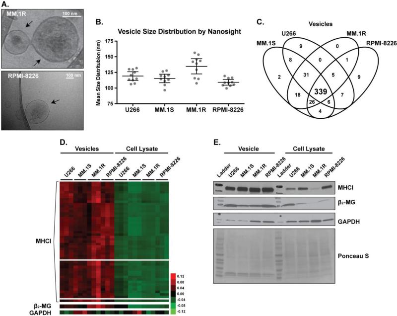
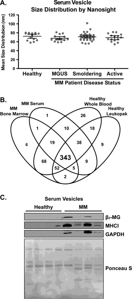
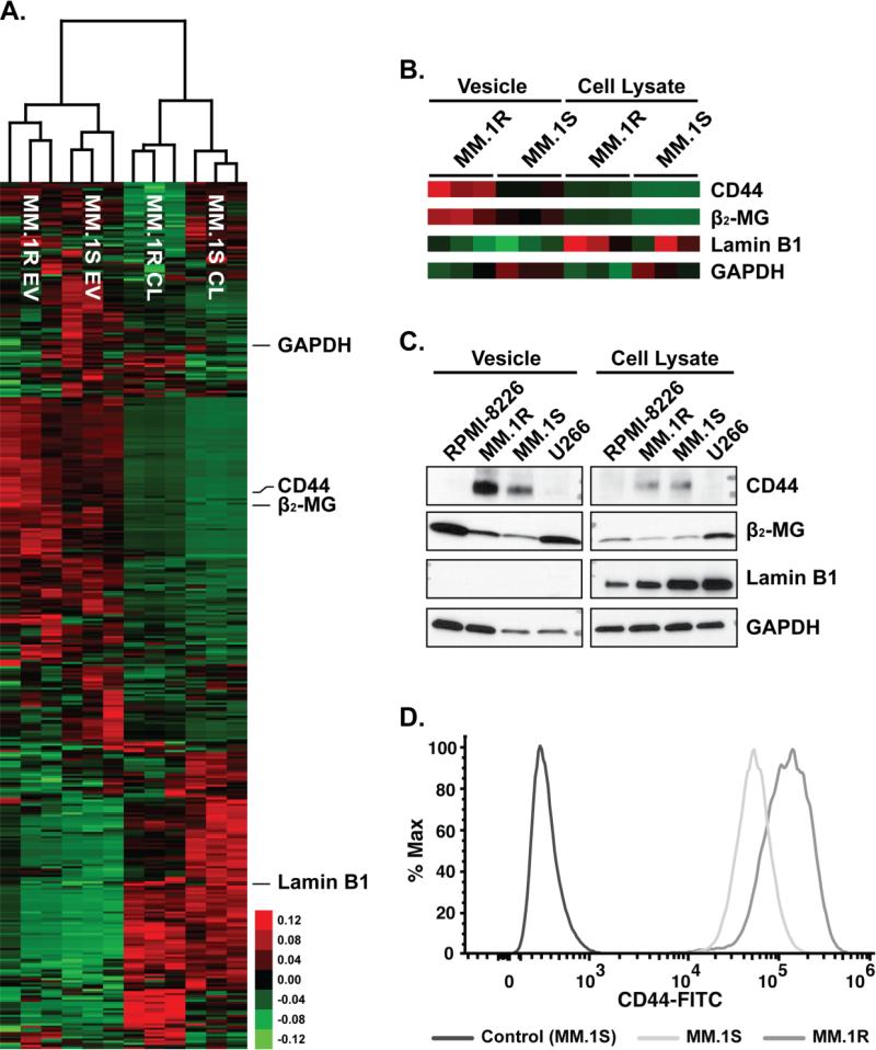
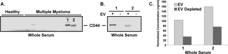
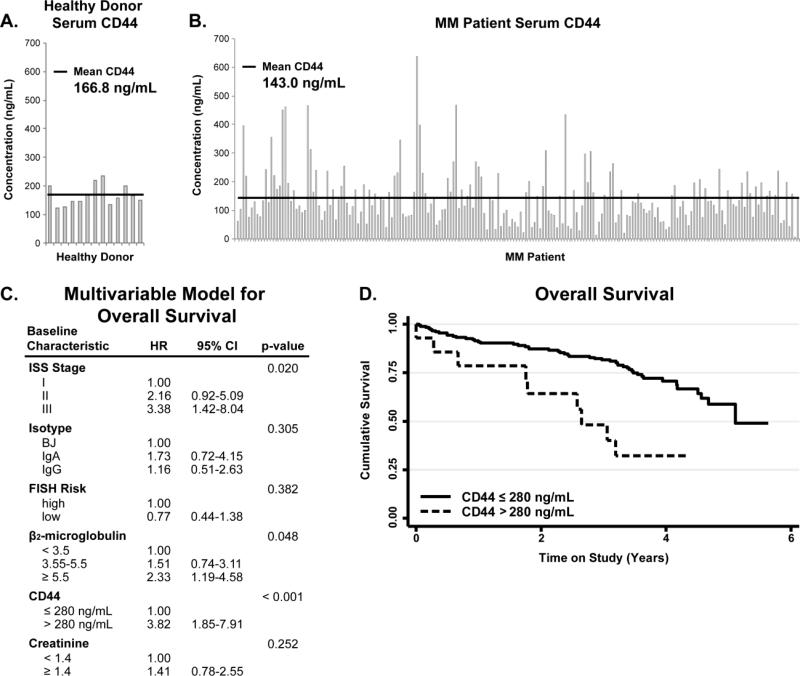
Multiple myeloma (MM) is a hematological malignancy of clonal plasma cells in the bone marrow (BM). The microenvironment plays a key role in MM cell survival and drug resistance through release of soluble factors, expression of adhesion molecules and release of extracellular vesicles (EVs). The aim of this manuscript is to use proteomic profiling of EVs as a tool to identify circulating tumor associated markers in MM patients. First, we characterized the EV protein content obtained from different MM cell lines. Then, we established differences in protein abundance among EVs isolated from MM patient serum and BM and the serum of healthy donors. These data show that the Major Histocompatibility Complex Class I is highly enriched in EVs of MM cell lines and MM patient's serum. Next, we show that CD44 is highly expressed in the EVs isolated from the corticosteroid resistant MM cell line, MM.1R. Furthermore, CD44 was found to be differentially expressed in EVs isolated from newly diagnosed MM patients. Finally through ELISA analysis, we establish the potential of serum CD44 as a predictive biomarker of overall survival. These results support the analysis of EVs as an easily accessible source for MM biomarkers.
Biological significance: Extracellular vesicles are becoming a research focus due to their roles in cancer cell biology such as immune evasion, therapeutic resistance, proliferation and metastases. While numerous studies of vesicle characterization and biology have been conducted in many cancer models, the role of EV in MM remains relatively unstudied. Here we found that EVs isolated from MM cells are enriched in MHC-1 antigen presenting complex and its binding protein β2-MG, this observation is compatible with the enhanced proteasome activity of MM cells compared to other cancers and the ability of functional MHC-1 to bind and present peptides, generated from protein degradation by the proteasome. Additionally, our experiments show that CD44 is particularly enriched in the EV fraction of corticosteroid resistant MM.1R cells and is differentially expressed in the EV fraction of MM patients. This is of high significance due to the established role of CD44 in adhesion of MM cells to BMSC and induction of IL-6, the primary cytokine for MM cell survival, secretion by the BMSC. Furthermore, ELISA assays for CD44 content from the serum of 254 newly diagnosed MM patients enrolled in a Phase 3 randomized trial show highly variable CD44 levels and those patients with >280 ng/mL serum CD44 showing a reduced overall survival time. These results suggest the potential use of CD44 as a prognostic biomarker in MM.
Keywords: Extracellular vesicles; Label-free relative quantitation; Multiple myeloma; Shotgun proteomics; cd44.
Copyright © 2016 Elsevier B.V. All rights reserved.
Figures
References
-
- American Cancer Society Cancer Facts & Figures. 2011:1–68. < http://www.cancer.org/acs/groups/content/@epidemiologysurveilance/docu m...>.
-
- Kuehl WM, Bergsagel PL. Multiple myeloma: evolving genetic events and host interactions. Nat Rev Cancer. 2002;2:175–187. - PubMed
-
- Vacca A, Ribatti D, Roccaro AM, Frigeri A, Dammacco F. Bone marrow angiogenesis in patients with active multiple myeloma. Semin Oncol. 2001;28:543–550. - PubMed
Publication types
MeSH terms
Substances
Grants and funding
LinkOut - more resources
Full Text Sources
Other Literature Sources
Medical
Research Materials
Miscellaneous
