. 2016 May;13(5):1172-1181.
doi: 10.1016/j.hrthm.2016.01.011. Epub 2016 Jan 13.

Cardiac Purkinje fibers and arrhythmias; The GK Moe Award Lecture 2015

Affiliations

Cardiac Purkinje fibers and arrhythmias; The GK Moe Award Lecture 2015

Penelope A Boyden et al. Heart Rhythm. 2016 May.

Abstract

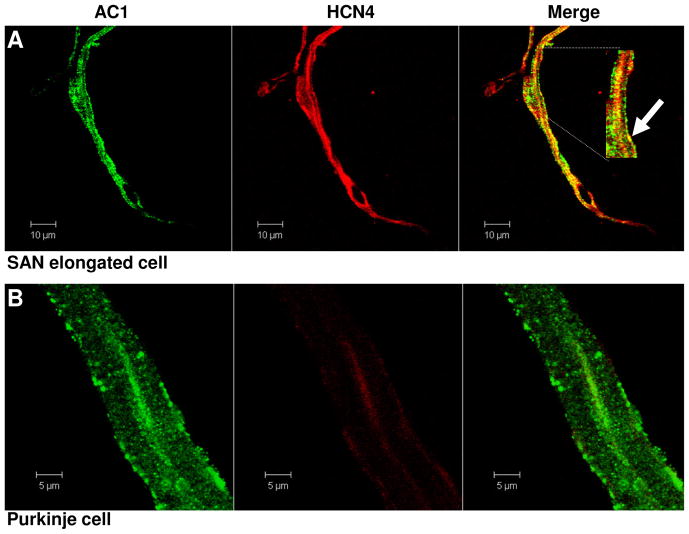
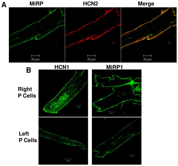
Purkinje fibers/cells continue to be a focus of arrhythmologists. Here we review several new ideas that have emerged in the literature and fold them into important new points. These points include the following: some proteins in Purkinje cells are specific to Purkinjes; pacemaker function in Purkinje may be similar to that of the sinus node cell; sink-source concerns about tracts/sheets of Purkinje fibers; role of Ito in arrhythmias; and genetic lesions in Purkinjes and their high impact on cardiac rhythm. Although new ideas about the remodeled Purkinje cell are not the focus of this review, one can easily imagine how Purkinjes and their function may be altered in diseased hearts.

Keywords: Arrhythmias; Calcium; Ion channels; Pacemaker activity; Purkinje.


Conflict of interest statement

There are no potential conflicts of interest.

Figures

Figure 1
Figure 1
A: Remarkably similar diagrams of the proximal left-sided conduction system as observed in 20 normal human hearts demonstrating the origin of the anterior, posterior and median fascicles from Syed et al. from [5]. B: Pig moderator band histology demonstrating conduction tissue (Purkinje fibers) lying within the band from Syed et al. [5]. C(a): A tissue block of India ink injected in the LV wall illustrating the most distal components of the conduction system. ED Endocardium, EP epicardium, PF intramyocardial and subendocardial Purkinje fibers. C(b): A tissue block of India ink injected left ventricular wall made transparent, showing the three dimensional (3D) intramyocardial network. Division = 1 mm; C(c): Detail of C(b); ED endocardium side, OF oblique intramyocardial communicating Purkinje fibers, VPF vertical intramyocardial Purkinje fibers. C(d): Histological section showing subendocardial fibers within India ink stained inside fibrous sheath. ED Endocardium, IK India ink, PF subendocardial Purkinje fibers. From De Almedia et al. [7]
Figure 2
Figure 2
A: A variety of phenotypes of canine single SAN cells: elongated (left), spider (right) and spindle (middle). Percentages of different phenotype cells from superior and inferior nodes are summarized below. E/S: elongated or spindle cell. B: Different characteristics of If in elongated/spindle (E/S) versus spider cells. Left: Activation kinetics differ significantly at indicated membrane potentials (*; Bonferoni). Right: Steady-state activation relations differ (ANOVA). C: Modification of cell aggregates of SAN AP parameters by isoprenaline 1 μM (Iso) emphasizing the effects on rate by the effects on EDD. C(a); Dot-plots of time course of action potential parameters during application of Iso (bar). C(b); Sample traces recorded in control and following perfusion with Iso, as indicated (arrows); corresponding takeoff potential (TOP) values plotted (filled squares, control; open squares, Iso). C(c); Cycle-by-cycle TOP vs rate plot (upper) and end diastolic depolarization(EDD)vs rate plot (lower) as from corresponding panels in A. From Bucchi et al. [21]
Figure 3
Figure 3
Immunofluorescent images of canine SAN(A) and Purkinje cells (B) co-stained with AC1 (Green) and HCN4 (Red). There was intense colocalization of AC1 and HCN4 in single SAN cells, however there was little immuno positive HCN4 protein in Purkinje cells.
Figure 4
Figure 4
A: Immunofluorescent images of canine Purkinje single cells co-stained with MiRP1 (Green) and HCN2 (Red), demonstrating that MiRP1 colocalizes with HCN2, the major isoform of the pacemaker channel in Purkinje cells B: Immunofluorescent images of the right and left ventricular Purkinje cells staining HCN1 (Left images) and MiRP1 (Right images). The staining of both proteins showed membrane immunofluorescence intensity, suggesting that HCN1 and MiRP1 colocalized on the membrane.
Figure 5
Figure 5
Representative If recorded from single canine SAN cells (L. Protas) (A) and Purkinje cells (B). Ivabradine significantly reduced If in SAN cells and decreased rate in both SAN and Purkinje cells, demonstrating that like SAN cells, the canine Purkinje fibers have ivabradine-sensitive rate effect (E. Sosunov, E. Anyukhovsky).
Figure 6
Figure 6
Role of Ito in EAD genesis in different species. A: The EAD distribution in the ICa,L and Ito conductance (GCa and Gto) space. The inactivation time constant of the Ito is the intermediate value (th0= 200 ms). EAD occurrence region is marked in grey. B: Single cell APs recorded in different species as indicated (a–d). The black and colored symbols mark the putative locations(including canine Purkinjes) of the APs in the distribution diagram (A). C: Summarized bar graphs showing the incidence of EADs within 20 Aps.. D: The same as (A), except that the inactivation time constant of the Ito is fast (th0= 20 ms), which is similar to that of canine ventricular myocytes. E: APs recorded from canine ventricular myocytes. The symbols mark the putative locations of the APs in the distribution diagram (C). From Zhao et al. [34].
Figure 7
Figure 7
Computational study showing that the number of cells exhibiting EAD or DAD matters. A(a):Schematic of 1D tissue, with central region of EAD/DAD susceptible cells demarcated in red, and unsusceptible cells in blue. A(b): Schematic of 2D tissue, with central elliptical region of EAD-/DAD-susceptible cells demarcated in red. A(c): Schematic of bricklike 2D tissue with fibroblasts (F) randomly interspersed at the ends (I) or sides (II) of the myocytes (M). B(a): Selected AP traces along a 1D cable, with the red traces indicating the EAD-susceptible cells in the central region, and the normal unsusceptible cells in black. The EAD failed to propagate with 69 susceptible cells in the central region (left), but propagated successfully with 70 susceptible cells (right). B(b): Same as for B(a), but with the central region exhibiting DAD-susceptible cells. The DAD failed to trigger an AP with 79 susceptible cells in the central region (left), but did so successfully with 80 susceptible cells (right). C: Number of contiguous susceptible myocytes required to trigger an EAD- or DAD-mediated PVC in simulated 2D tissue with lateral fibrosis. Fibroblasts were randomly interspersed exclusively along the sides of myocytes throughout the entire tissue, with EAD- or DAD-susceptible myocytes only in the central region. As the fibroblast/myocyte (FM) ratio increased, the required size of the central region (and hence the number of susceptible cells in the central region) progressively decreased, approaching the 1D case at the maximum FM ratio of 5, above which transverse propagation failed. From Xie et al. [39]
Figure 8
Figure 8
Abnormal repolarization and ventricular fibrillation. A: Presenting ECG of proband with right bundle branch block; J point elevation (arrows) <2mV in leads V1-V3 thus not consistent with Brugada pattern. B: Ectopy with short-coupled PVCs. C: Short-coupled PVC initiates VF requiring ICD shock in the presence of quinidine. Over 16 months, proband had 168 appropriate ICD discharges for VF (~8.6 per month). D: DPP6-T H332R displays increased Kv4.3 binding relative to DPP6-T. D(a) and D(b); Co-immunoprecipitations of Kv4.3 and DPP6-T from human left ventricle lysates. D(c); Purified GST-Kv4.3, but not GST alone associates with DPP6-T in pull-down experiments from detergent-soluble human left ventricle lysates. D(d) and D(e) Purified GST-Kv4.3, but not GST associates with radiolabelled DPP6-T and DPP6-T H332R. However, GST-Kv4.3 shows over 2 fold increase in relative binding activity for DPP6-T H332R versus DPP6-T when corrected for input and protein loading. E: DPP6-T+ Kv4.3 displays augmented Ito compared with DPP6+ Kv4.3. E(a) and E(b);DPP6-T H332R +Kv4.3 shows ~7 fold increase in peak Ito compared with WT DPP6-T. From Sturm et al. [40]

Similar articles

Cited by

References

    1. MENDEZ C, MUELLER WJ, MERIDETH J, MOE GK. Interaction of Transmembrane Potentials in Canine Purkinje Fibers and at Purkinje Fiber-Muscle Junctions. Circ Res. 1969;24:361–72. - PubMed
    1. Singer DH, Lazzara R, Hoffman BF. Interrelationships between automaticity and conduction in Purkinje fibers. Circ Res. 1967;21:537–58. - PubMed
    1. Robinson RB, Boyden PA, Hoffman BF, Hewett KW. The electrical restitution process in dispersed canine cardiac Purkinje and ventricular cells. Am J Physiol. 1987;253:H1018–H1025. - PubMed
    1. Boyden PA, Hirose M, Dun W. Cardiac Purkinje cells. Heart Rhythm. 2010;7:127–35. - PubMed
    1. Syed FF, Hai JJ, Lachman N, DeSimone CV, Asirvatham SJ. The infrahisian conduction system and endocavitary cardiac structures: relevance for the invasive electrophysiologist. J Interv Card Electrophysiol. 2014;39:45–56. - PMC - PubMed

MeSH terms