Review
. 2016 May;15(5):348-66.
doi: 10.1038/nrd.2015.6. Epub 2016 Jan 18.

Regulated necrosis: disease relevance and therapeutic opportunities

Affiliations
Review

Regulated necrosis: disease relevance and therapeutic opportunities

Marcus Conrad et al. Nat Rev Drug Discov. 2016 May.

Abstract

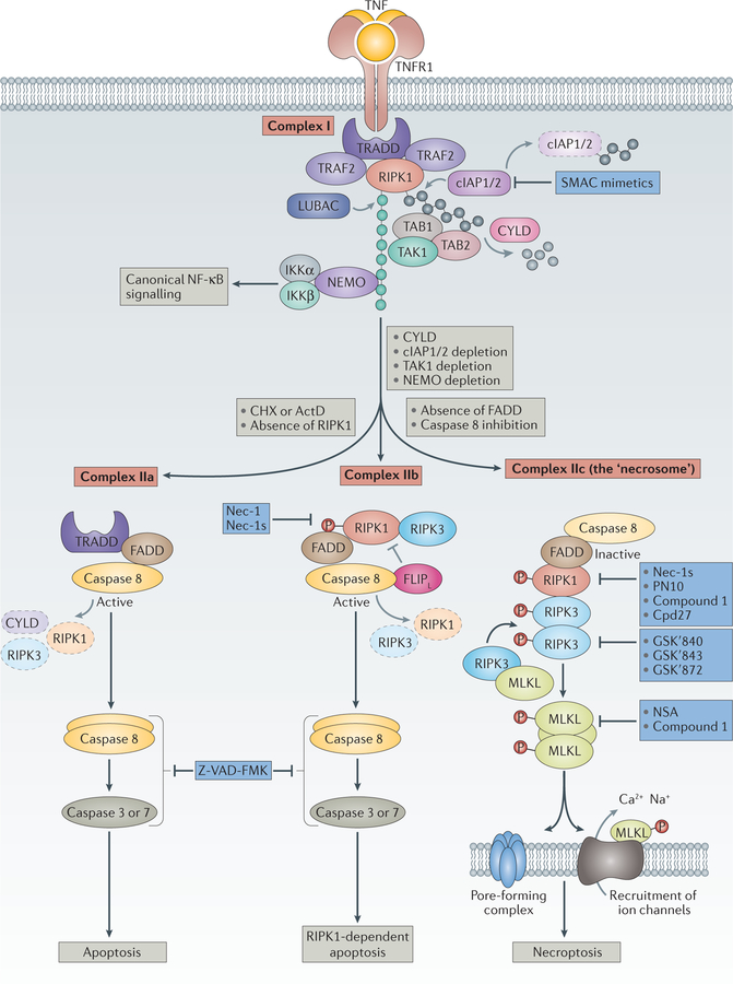
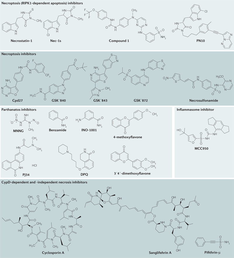
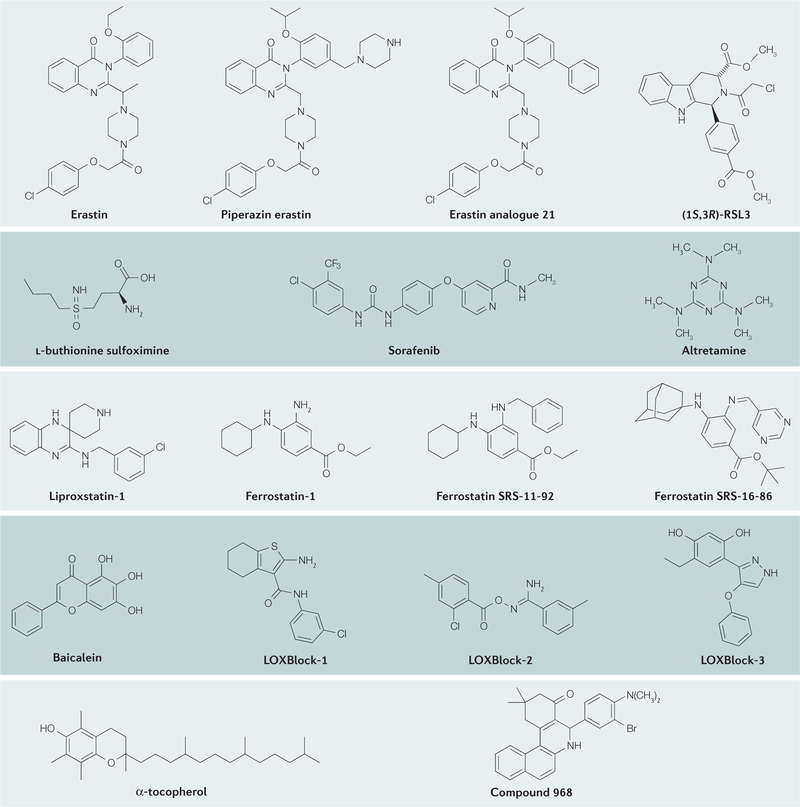
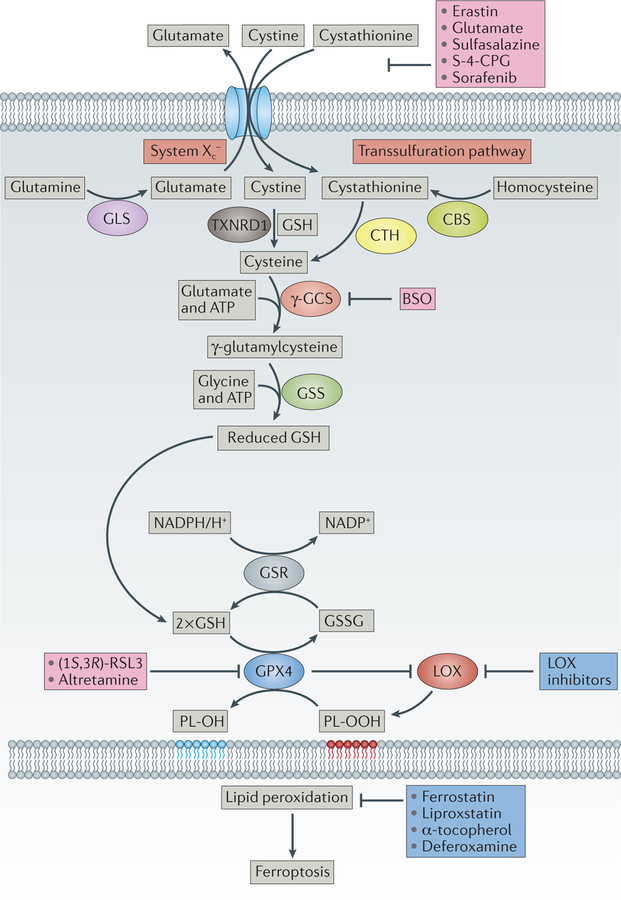
The discovery of regulated cell death presents tantalizing possibilities for gaining control over the life-death decisions made by cells in disease. Although apoptosis has been the focus of drug discovery for many years, recent research has identified regulatory mechanisms and signalling pathways for previously unrecognized, regulated necrotic cell death routines. Distinct critical nodes have been characterized for some of these alternative cell death routines, whereas other cell death routines are just beginning to be unravelled. In this Review, we describe forms of regulated necrotic cell death, including necroptosis, the emerging cell death modality of ferroptosis (and the related oxytosis) and the less well comprehended parthanatos and cyclophilin D-mediated necrosis. We focus on small molecules, proteins and pathways that can induce and inhibit these non-apoptotic forms of cell death, and discuss strategies for translating this understanding into new therapeutics for certain disease contexts.


Conflict of interest statement

Competing interests statement

The authors declare competing interests: see Web version for details.

Figures

Figure 1
Figure 1. The main signalling events downstream of TNFR activation.
Binding of tumour necrosis factor (TNF) to its cognate receptor TNFR1 triggers the assembly of complex I, which comprises TNFR1, TNFR1-associated death domain (TRADD), receptor-interacting serine/threonine-protein kinase 1 (RIPK1), TNFR-associated factor 2 (TRAF2), cellular inhibitor of apoptosis protein 1/2 (cIAP1/2), and linear ubiquitin chain assembly complex (LUBAC),,. Complex I provides the platform for Lys63-linked ubiquitylation (grey circles) or linear ubiquitylation (green circles) of RIPK1 by cIAP1/2 or LUBAC, respectively. This ubiquitylation is implicated in the decision between nuclear factor-κB (NF-κB) or survival signalling and cell death signalling. Ubiquitylation leads to the recruitment of other factors such as transforming growth factor-β (TGFβ)-activated kinase (TAK1), TAK1-binding protein 1 (TAB1) and TAB2, or NF-κB essential modulator (NEMO) and the inhibitor of the NF-κB kinase-α (IKKα)–IKKβ complex, usually triggering canonical NF-κB signalling. In the presence of cycloheximide (CHX), a translational inhibitor, TNF stimulation leads to the formation of a cytosolic complex IIa in which RIPK1 disappears, whereas TRADD and FAS-associated death domain (FADD) interaction leads to the activation of caspase 8 and effector caspases such as caspase 3 and caspase 7 and apoptotic cell death. With cIAP1/2 inhibitors (SMAC (second mitochondria-derived activator of caspase) mimetics), knockdown of cIAPs,– or inhibition or depletion of TAK1 or NEMO, complex IIb is formed. Complex IIb consists of RIPK1, RIPK3, FADD and caspase 8, and favours RIPK1-kinase-activity-dependent apoptosis. Heteromeric pro-caspase 8–FLICE-like inhibitory protein long isoform (FLIPL) inhibits necroptosis, probably by cleaving RIPK1, RIPK3 and cylindromatosis (CYLD). With sufficient expression of RIPK3 and concomitant inhibition or reduced expression of pro-caspase 8 and FLIPL (REF. 26), complex IIc (also known as the necrosome) is formed,,. The formation of complex IIc entails the association of RIPK1 and RIPK3 followed by a series of auto-and transphosphorylation events of RIPK1 and RIPK3. Activated RIPK3 phosphorylates and recruits mixed lineage kinase domain-like protein (MLKL),, eventually leading to the formation of a supramolecular protein complex at the plasma membrane and necroptosis–,. SMAC mimetics are being developed to impair survival signalling and to sensitize cells to the triggering of cell death in the context of tumour treatment. Z-VAD-FMK is a bona fide pan-caspase inhibitor. Necrostatin-1 (Nec-1) and the more-specific Nec-1s, Cpd27 (REF. 41) and, more recently, a hybrid molecule consisting of Nec-1s and ponatinib called PN10 (REF. 42) all inhibit the kinase activity of RIPK1 and thus inhibit necroptotic signalling. Additional necroptosis inhibitors include the RIPK3 inhibitors GSKʹ840, GSKʹ843 and GSKʹ872 (REF. 33), as well as the MLKL inhibitors necrosulfonamide (NSA) and compound 1 (REF. 34). However, the specificity of these MLKL inhibitors is not restricted to inhibition of MLKL, and their efficacy is probably also due to effects on other steps in the necroptosis pathway. ActD, actinomycin D.
Figure 2
Figure 2. Chemical structures of inhibitors of non-apoptotic cell death.
The mechanisms of action, key functions and references for these inhibitors are provided in TABLE 1. CypD, cyclophilin D; RIPK1, receptor-interacting serine/threonine kinase 1.
Figure 3
Figure 3. Chemical structures of ferroptosis inducers and inhibitors.
The mechanisms of action, key functions and references for these inhibitors are provided in TABLE 1.
Figure 4
Figure 4. Upstream events in the control of ferroptosis.
The heterodimeric cystine–glutamate antiporter system Xc exchanges one molecule of extracellular cystine (or cystathionine) for one molecule of intracellular glutamate57. Once taken up by cells, cystine is reduced by reduced glutathione (GSH) or thioredoxin reductase 1 (TXNRD1) to cysteine, which is subsequently used for protein and GSH synthesis. Cysteine might also be provided by the transsulfuration pathway. GSH is synthesized from cysteine, glutamate and glycine in two consecutive steps by γ-glutamylcysteine synthetase (γ-GCS) and glutathione synthase (GSS) at the expense of two molecules of ATP. Glutathione peroxidase 4 (GPX4) is one of the central upstream regulators of ferroptosis: it prevents lipoxygenase (LOX) overactivation and lipid peroxidation. GPX4 preferentially reduces phospholipid hydroperoxide (PL-OOH) to its corresponding alcohol phospholipid hydroxide (PL-OH), using two molecules of GSH. Oxidized glutathione (GSSG) is then recycled back by glutathione reductase (GSR) using electrons from NADPH/H+. Concerted action of this pathway is essential to control the formation of oxidized phospholipids. A series of ferroptosis inducers have been developed and characterized that interfere at the different upstream events, either inhibiting cystine uptake (for example, erastin, glutamate, sulfasalazine, (S)-4-carboxyphenylglycine (S-4-CPG) and sorafenib), GSH biosynthesis (L-buthionine sulfoximine (BSO)) or GPX4 activity ((1S, 3R)-RSL3 (REF. 60) or altretamine). Ferrostatins, liproxstatin, α-tocopherol and the iron chelator deferoxamine inhibit lipid peroxidation and signalling events downstream of GPX4 function. CBS, cystathionine β-synthase; GLS, glutaminase; CTH, cystathionase.
Figure 5
Figure 5. Key signalling events in parthanatos.
DNA-damaging agents trigger poly(ADP-ribose) (PAR) polymerase 1 (PARP1) activation, the DNA-damage response and the repair response. In neurons, sustained stimulation of N-methyl-D-aspartate (NMDA) receptors (for instance, following stroke or traumatic brain injury) also leads to parthanatos, through cellular increases in Ca2+ levels, activation of neuronal nitric oxide synthase (nNOS), increased superoxide production and generation of highly toxic peroxynitrite (ONOO) ions. PARP-mediated PARylation (blue circles) of target proteins, and of PARP1 itself, leads to recruitment of factors required for DNA repair (not shown). The increase in PARylated proteins is counterbalanced by poly(ADP-ribose) glycohydrolase (PARG), which cleaves glycosidic bonds in PAR polymers on target proteins and thereby regulates PAR length and releases PAR polymers. ADP-ribosyl-acceptor hydrolase 3 (ARH3) further degrades PAR polymers. Excess DNA damage can elicit parthanatos by inducing PARylation of apoptosis-inducing factor (AIF) and relocation of AIF into the nucleus, as well as PARylation of hexokinase (HK), which impairs its glycolytic activity. AIF translocation to the nucleus is associated with large-scale fragmentation of DNA and cell death. Inhibitors of parthanatos include the PARP1 inhibitors benzamide and its derivatives, such as INO-1001, methoxyflavones, PJ34 (REF. 180) and DPQ (3,4-dihydro-5-[4-(1-piperidinyl)butoxyl]-1(2H)-isoquinolinone). MNNG, N-methyl-Nʹ-nitro-N-nitrosoguanidine; NAM, nicotinamide; NO, nitric oxide; O2, superoxide anion; ROS, reactive oxygen species.

Similar articles

Cited by

References

    1. Laster SM, Wood JG & Gooding LR Tumor necrosis factor can induce both apoptic and necrotic forms of cell lysis. J. Immunol 141, 2629–2634 (1988). - PubMed
    1. Vanden Berghe T, Linkermann A, Jouan-Lanhouet S, Walczak H & Vandenabeele P Regulated necrosis: the expanding network of non-apoptotic cell death pathways. Nat. Rev. Mol. Cell Biol 15, 135–147 (2014). - PubMed
    1. Green DR & Levine B To be or not to be? How selective autophagy and cell death govern cell fate. Cell 157, 65–75 (2014). - PMC - PubMed
    1. Galluzzi L, Pietrocola F, Levine B & Kroemer G Metabolic control of autophagy. Cell 159, 1263–1276 (2014). - PMC - PubMed
    1. Brinkmann V & Zychlinsky A Neutrophil extracellular traps: is immunity the second function of chromatin? J. Cell Biol 198, 773–783 (2012). - PMC - PubMed

Substances