. 2016 Jan 14;7(1):e2051.
doi: 10.1038/cddis.2015.386.

HSP90 activity is required for MLKL oligomerisation and membrane translocation and the induction of necroptotic cell death

Affiliations

HSP90 activity is required for MLKL oligomerisation and membrane translocation and the induction of necroptotic cell death

A V Jacobsen et al. Cell Death Dis. .

Abstract

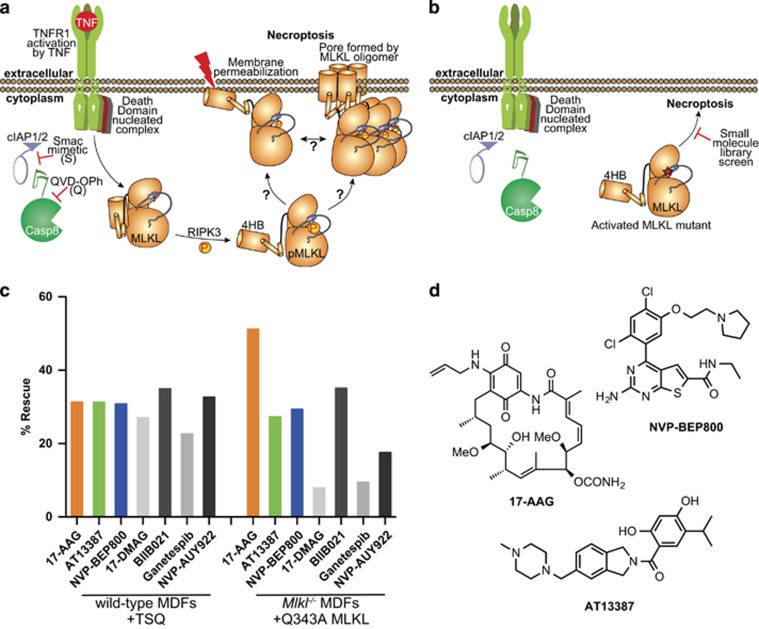
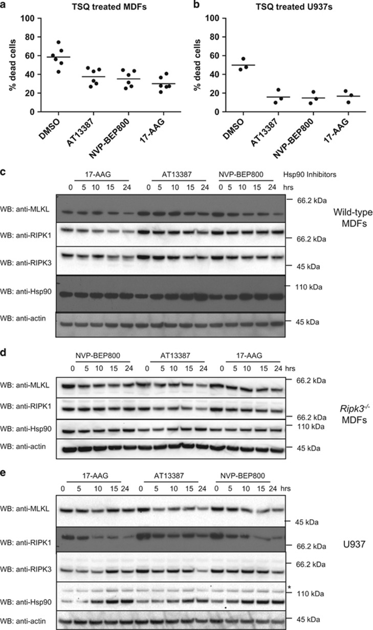
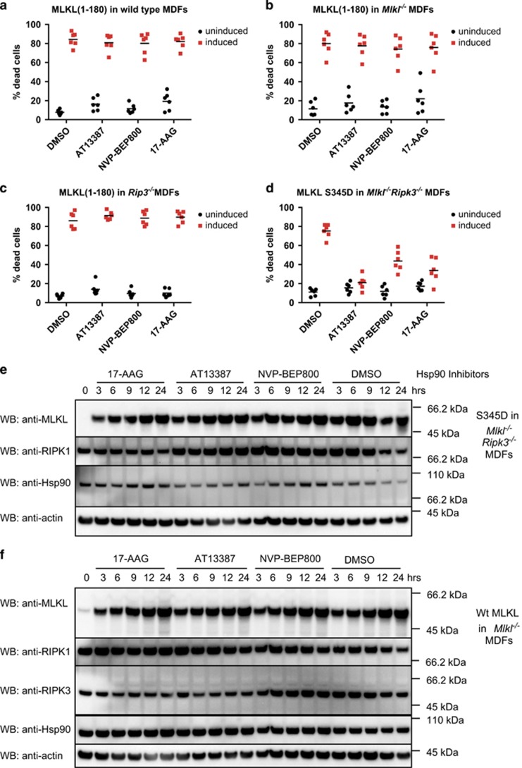
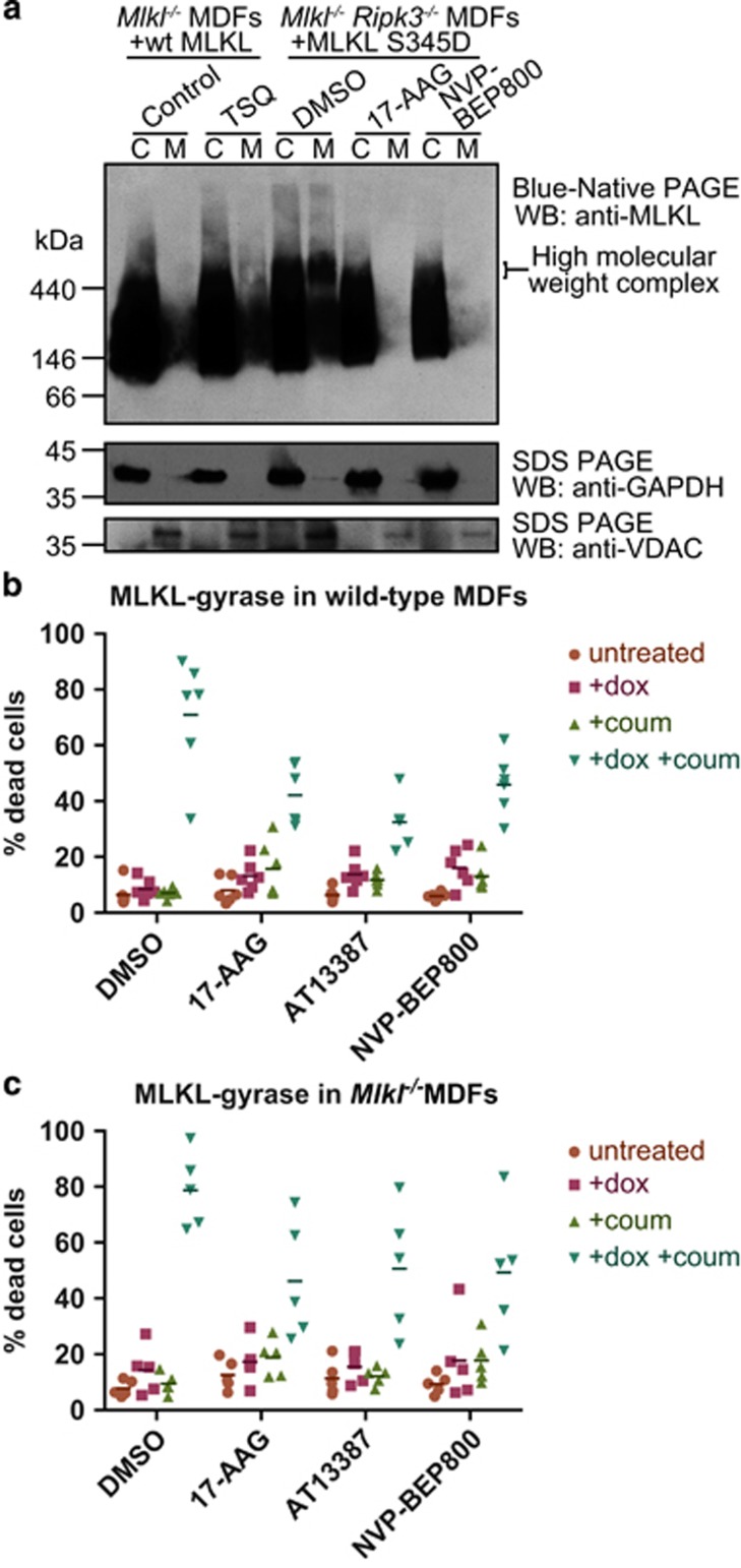
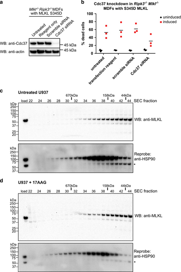
Necroptosis is a caspase-independent form of regulated cell death that has been implicated in the development of a range of inflammatory, autoimmune and neurodegenerative diseases. The pseudokinase, Mixed Lineage Kinase Domain-Like (MLKL), is the most terminal known obligatory effector in the necroptosis pathway, and is activated following phosphorylation by Receptor Interacting Protein Kinase-3 (RIPK3). Activated MLKL translocates to membranes, leading to membrane destabilisation and subsequent cell death. However, the molecular interactions governing the processes downstream of RIPK3 activation remain poorly defined. Using a phenotypic screen, we identified seven heat-shock protein 90 (HSP90) inhibitors that inhibited necroptosis in both wild-type fibroblasts and fibroblasts expressing an activated mutant of MLKL. We observed a modest reduction in MLKL protein levels in human and murine cells following HSP90 inhibition, which was only apparent after 15 h of treatment. The delayed reduction in MLKL protein abundance was unlikely to completely account for defective necroptosis, and, consistent with this, we also found inhibition of HSP90 blocked membrane translocation of activated MLKL. Together, these findings implicate HSP90 as a modulator of necroptosis at the level of MLKL, a function that complements HSP90's previously demonstrated modulation of the upstream necroptosis effector kinases, RIPK1 and RIPK3.


Conflict of interest statement

JS is a member of the Scientific Advisory Board of TetraLogic Pharmaceuticals. GL and JMM lead a programme funded by Catalyst Therapeutics Pty Ltd and the Walter and Eliza Hall Institute to develop necroptosis inhibitors.

Figures

Figure 1
Figure 1
Hsp90 inhibitors can block necroptosis downstream of MLKL activation. (a) A schematic of the necroptosis pathway. TNF (T) stimulates the TNFR1; cIAP1/2 activity is blocked with Smac mimetic (S); and the pan-caspase inhibitor, QVD-OPh (Q), blocks caspase-8 activity. This leads to RIPK3 activation and subsequent phosphorylation and activation of MLKL. (b) The activated MLKL mutant Q343A initiates cell death in the absence of TSQ stimulation or RIPK3 activation enabling a screen for inhibitors of necroptosis downstream of MLKL activation. (c) Cells were pretreated for 2 h with a library of 438 compounds with annotated mechanisms of action at 1 μM, then necroptosis was stimulated in wild-type MDFs with TSQ and expression of Q343A MLKL was induced in Mlkl−/− MDFs with doxycycline. Percentage rescue was determined using a CellTiter Glo assay, and normalised against TSQ/doxycycline stimulation and DMSO treatment. The data represent the HSP90 hits from each screen, and are the average of two technical replicates. (d) Chemical structures of the three compounds selected for further analysis
Figure 2
Figure 2
MLKL levels are modestly reduced by Hsp90 inhibition. (a) MDFs were pretreated for 1 h with AT13387 (1 μM), NVP-BEP800 (125 nM), 17-AAG (250 nM) or DMSO, then necroptosis was induced with TSQ. After 24 h, propidium iodide (PI) uptake was measured using flow cytometry. Each data point represents results from one of three independent cell lines tested in two experiments and solid bar indicates average (n=6). (b) U937 cells were pretreated for 1 h with AT13387 (2 μM), NVP-BEP800 (1 μM), 17-AAG (500 nM) or DMSO, then necroptosis was induced with TSQ. After 24 h, PI uptake was measured using flow cytometry. Each data point represents results from the U937 cell line tested in three independent experiments and solid bar indicates average (n=3). (c and d) Wild-type (c) or Ripk3−/− (d) MDFs were treated with AT13387 (1 μM), NVP-BEP800 (125 nM) or 17-AAG (250 nM) for the indicated times, then cell lysates analysed with western blotting using the indicated antibodies. Data are representative of three independent experiments. (e) U937 cells were treated with AT13387 (2 μM), NVP-BEP800 (1 μM) or 17-AAG (500 nM) over a 24-h time course, then cell lysates analysed with western blotting using the indicated antibodies. Data are representative of three independent experiments. *Represents non-specific band at ~110 kDa
Figure 3
Figure 3
Hsp90 inhibition affects MLKL activity. (ac) Wild-type (a), Mlkl−/− (b) or Ripk3−/− (c) MDFs were pretreated for 1 h with AT13387 (1 μM), NVP-BEP800 (125 nM), 17-AAG (250 nM) or DMSO, then expression of MLKL(1–180) was induced using 50 ng/ml doxycycline. After 24 h, PI uptake was measured using flow cytometry. Two experiments were performed using three independent cell lines (n=6). (d) Mlkl−/− Ripk3−/− MDFs were pretreated with HSP90 inhibitors as described above, then expression of MLKL S345D was induced using 50 ng/ml doxycycline. After 24 h, PI uptake was measured using flow cytometry. Two experiments were performed using three independent cell lines (n=6). (e and f) Cells were pretreated for 1 h with AT13387 (1 μM), NVP-BEP800 (125 nM), 17-AAG (250 nM) or DMSO, then expression of MLKL S345D in Mlkl−/−Ripk3−/− double knockout MDFs (e) or endogenous MLKL in Mlkl−/− MDFs (f) was induced using 50 ng/ml doxycycline. Treatment was performed over 24 h at the times shown, then cell lysates analysed with western blotting using the indicated antibodies. Data are representative of three independent experiments
Figure 4
Figure 4
HSP90 is required for oligomerisation and membrane translocation of MLKL. (a) Mlkl−/− Ripk3−/− MDFs were pretreated for 1 h with NVP-BEP800 (125 nM), 17-AAG (250 nM) or DMSO then expression of MLKL S345D was induced using 50 ng/ml doxycycline. Cells were harvested when ~40% of DMSO-treated cells were dead (~7 h after induction). Expression of wild-type MLKL in Mlkl−/− MDFs was induced using 50 ng/ml doxycycline for 3 h, then cells were either left untreated (Control) or treated with TSQ to induce necroptosis. Cells were harvested when cell death was ~40% in TSQ-treated cells (~5 h after TSQ treatment). Cell lysates were then separated into cytoplasmic (C) or membrane (M) fractions, and separated using Blue Native PAGE. Levels of MLKL were analysed using western blotting. Data are representative of three independent experiments using two biologically independent cell lines. (b and c) Expression of wild-type full-length MLKL fused via its C-terminus to DNA gyraseB was expressed in wild-type (b) or Mlkl−/− (c) MDFs using 50 ng/ml doxycycline (+dox) and were dimerised via treatment with 700 nM coumermycin (+coum). Cells were pretreated for 1 h with AT13387 (1 μM), NVP-BEP800 (125 nM), 17-AAG (250 nM) or DMSO, before MLKL-gyrase expression was induced with dox. After 24 h, propidium iodide (PI) uptake was measured using flow cytometry. Each data point represents results from one of three independent cell lines tested in two experiments, and solid bar indicates mean (n=6 for wild-type MDFs; n=5 for Mlkl−/− MDFs)
Figure 5
Figure 5
MLKL transiently interacts with HSP90 via the Cdc37 co-chaperone. (a and b) Mlkl−/− Ripk3−/− MDFs stably transduced with a lentiviral construct encoding S345D MLKL were untreated, treated with transfection reagent only, or transfected with scrambled or Cdc37 siRNA pools. Cdc37 knockdown was observed in Cdc37 siRNA-treated cells relative to untreated, transfection reagent and scrambled siRNA-treated controls by western blot (a). Only Cdc37 siRNA knockdown conferred protection from S345D MLKL-mediated death (b). (c and d) Lysates of U937 cells incubated with DMSO (c) or 17-AAG (500 nM, d) were resolved by Superose-6 10/300 size-exclusion chromatography (SEC). Fractions were subjected to SDS-PAGE and western blotted for MLKL (upper panels) before reprobing for HSP90 (lower; * corresponds to residual signal from MLKL blots). The SEC fraction number is shown above the blots along with the elution position of molecular weight standards

References

    1. Silke J, Rickard JA, Gerlic M. The diverse role of RIP kinases in necroptosis and inflammation. Nat Immunol 2015; 16: 689–697. - PubMed
    1. Cho YS, Challa S, Moquin D, Genga R, Ray TD, Guildford M et al. Phosphorylation-driven assembly of the RIP1-RIP3 complex regulates programmed necrosis and virus-induced inflammation. Cell 2009; 137: 1112–1123. - PMC - PubMed
    1. Upton JW, Kaiser WJ, Mocarski ES. Virus inhibition of RIP3-dependent necrosis. Cell Host Microbe 2010; 7: 302–313. - PMC - PubMed
    1. Fan H, Liu F, Dong G, Ren D, Xu Y, Dou J et al. Activation-induced necroptosis contributes to B-cell lymphopenia in active systemic lupus erythematosus. Cell Death Dis 2014; 5: e1416. - PMC - PubMed
    1. Gunther C, Martini E, Wittkopf N, Amann K, Weigmann B, Neumann H et al. Caspase-8 regulates TNF-alpha-induced epithelial necroptosis and terminal ileitis. Nature 2011; 477: 335–339. - PMC - PubMed

Publication types