. 2016 Jan 14;7(1):e2059.
doi: 10.1038/cddis.2015.361.

Effect of hydroxychloroquine and characterization of autophagy in a mouse model of endometriosis

Affiliations

Effect of hydroxychloroquine and characterization of autophagy in a mouse model of endometriosis

A Ruiz et al. Cell Death Dis. .

Abstract

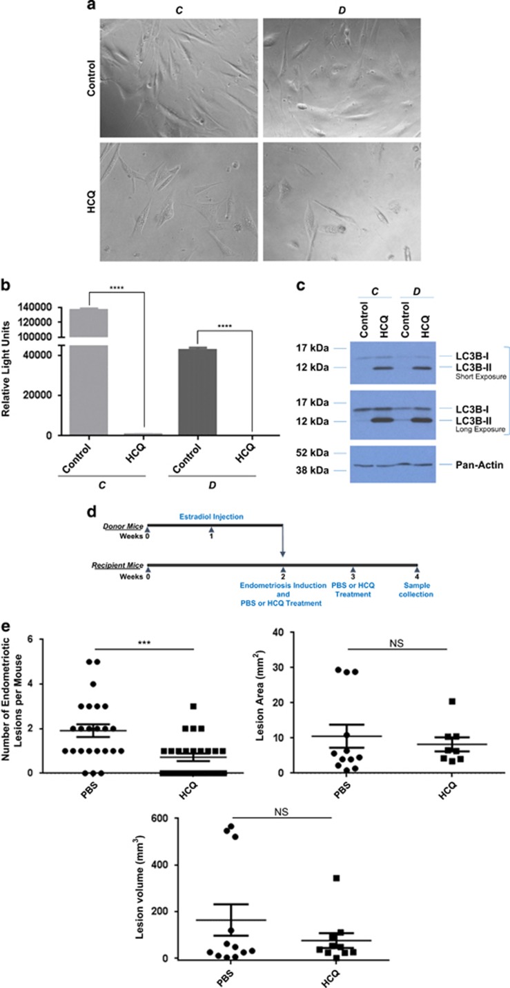
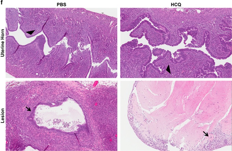
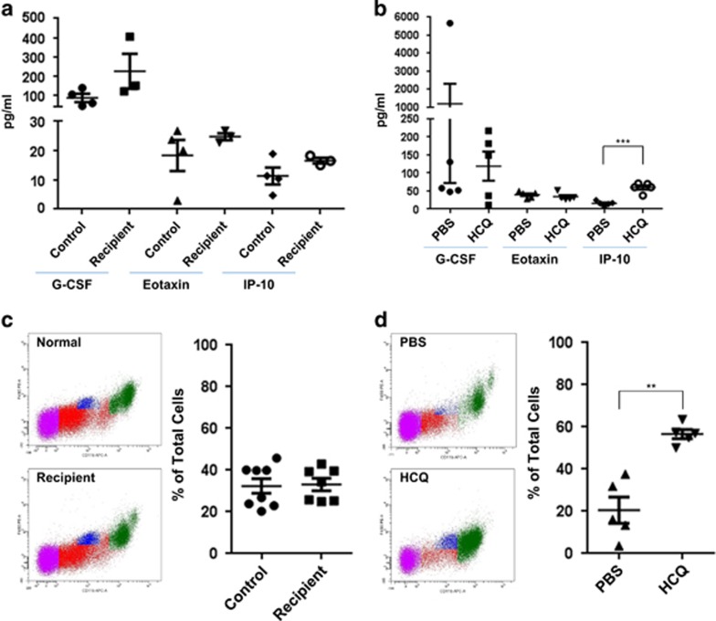
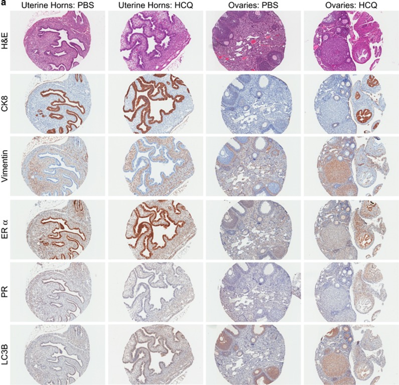
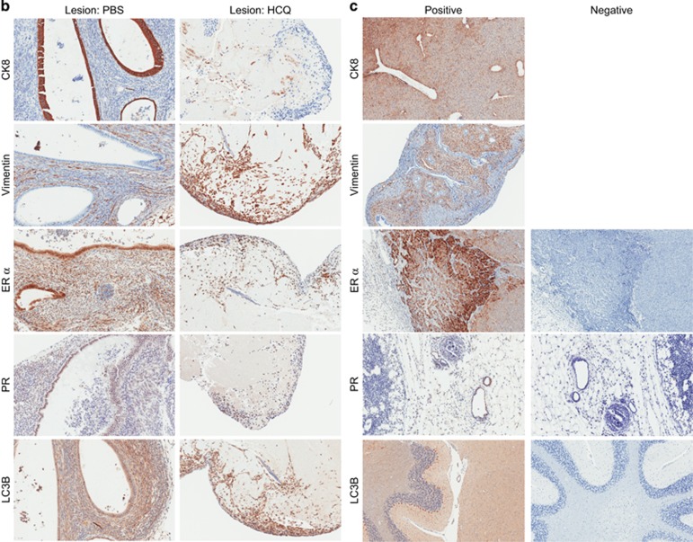
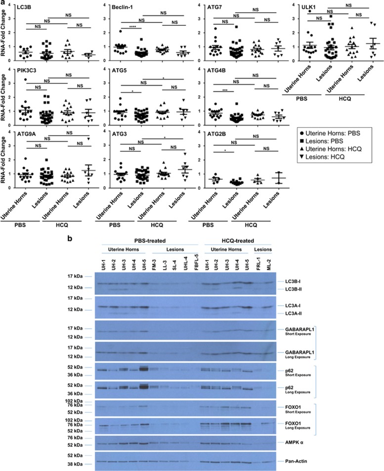
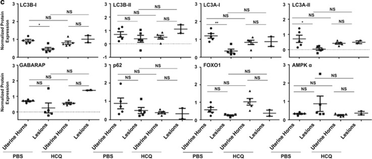
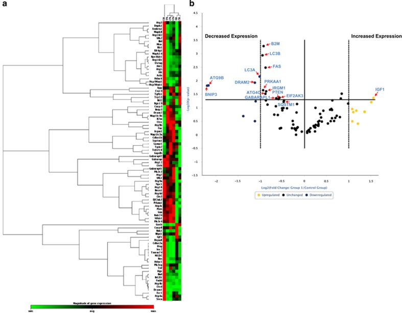
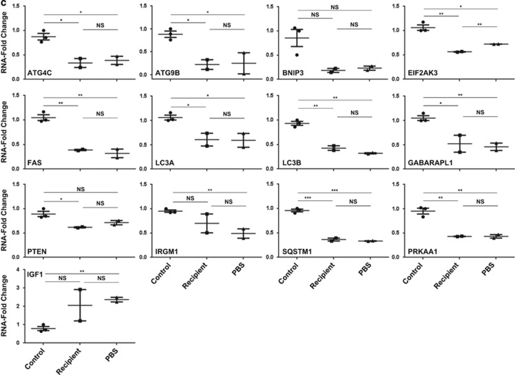
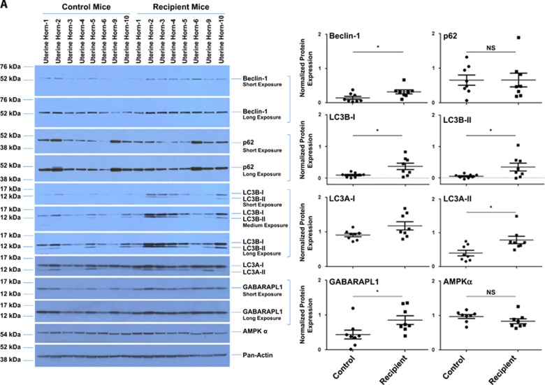
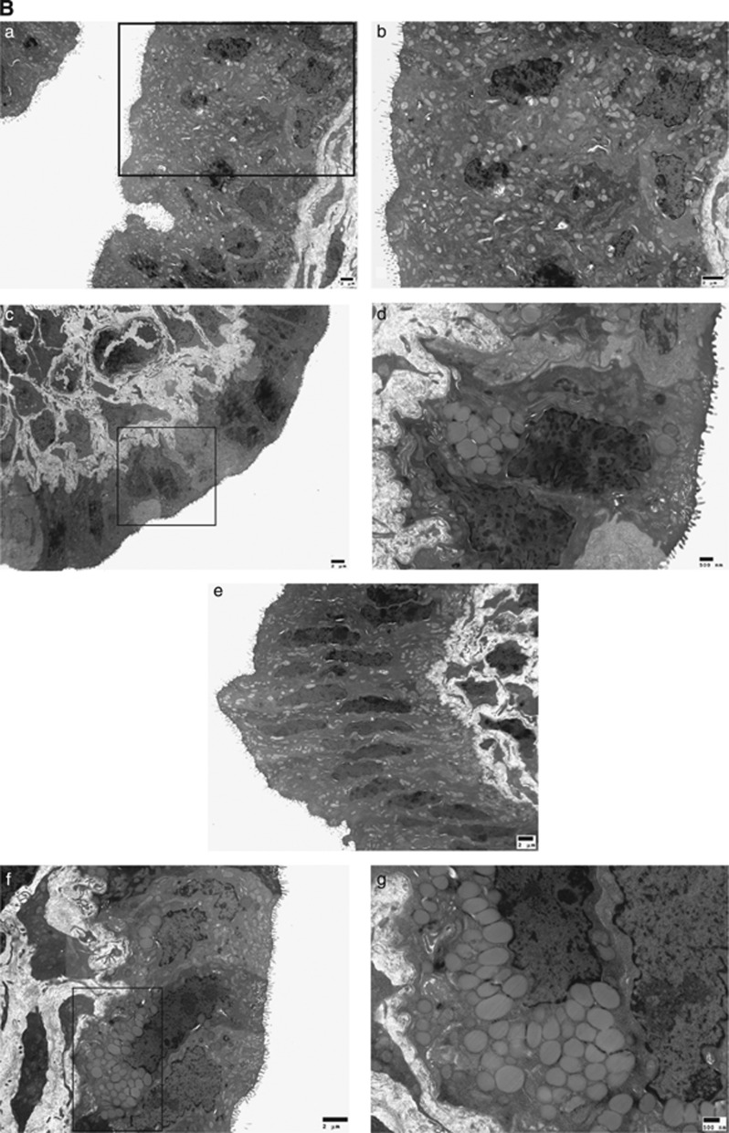
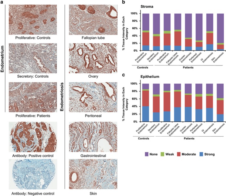
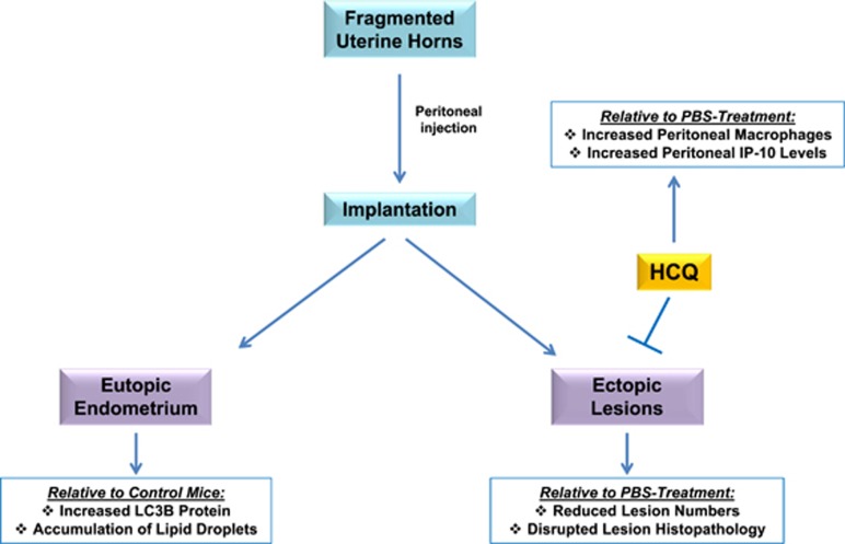
In endometriosis, the increased survival potential of shed endometrial cells (which normally undergo anoikis) is suggested to promote lesion development. One mechanism that may alter anoikis is autophagy. Using an autophagic flux inhibitor hydroxychloroquine (HCQ), we identified that it reduces the in vitro survival capacity of human endometriotic and endometrial T-HESC cells. We also identified that HCQ could decrease lesion numbers and disrupt lesion histopathology, as well as increase the levels of peritoneal macrophages and the IP-10 (10 kDa interferon-γ-induced protein) chemokine in a mouse model of endometriosis. We noted that RNA levels of a subset of autophagic markers were reduced in lesions relative to uterine horns from endometriosis-induced (untreated) mice. In addition, the RNA levels of autophagic markers were decreased in uterine horns of endometriosis-induced mice compared with those from controls. However, we noted that protein expression of LC3B (microtubule-associated protein 1 light-chain 3β; an autophagic marker) was increased in uterine horns of endometriosis-induced mice compared with uterine horns of controls. By immunohistochemical staining of a human endometriosis-focused tissue microarray, we observed LC3B expression predominantly in epithelial relative to stromal cells in both eutopic and ectopic endometria. Via transmission electron microscopy, cells from eutopic endometria of endometriosis-induced mice contained more lipid droplets (rather than autophagosomes) compared with uterine horns from controls. Collectively, our findings indicate that the autophagic pathway is dysregulated in both ectopic and eutopic endometrium in a murine model of endometriosis and that HCQ has potential as a therapeutic agent for women afflicted with endometriosis.


Conflict of interest statement

A provisional patent application on autophagy and endometriosis has been submitted” (MN, IF). All other co-authors declare no conflict of interest.

Figures

Figure 1
Figure 1
HCQ reduces endometriotic cell survival as well as lesion number and histopathology in a mouse model of endometriosis. (a) Representative images of life-extended endometriotic cells using human endometriotic cells derived from two different lesion types: ‘C' and ‘D' treated for 18 h with 25 μM HCQ. (b) Cell survival of life-extended endometriotic cells treated with 25 μM HCQ for 5 days was assessed by CellTiter-glo and measuring luminescence. (c) Cell lysate from life-extended endometriotic cells treated with 25 μM HCQ for 18 h were analyzed by western blotting using the indicated antibodies. Three independent experiments were performed. (d) Schematic representation of the experimental design. Mice were intraperitoneally injected with β-estradiol at 6 weeks of age. After 1 week, these mice were killed and their uterine horns were removed, minced, and injected into the peritoneal cavity of the same-age mice (recipients at 7 weeks of age). The same day of endometriosis induction, mice received an intraperitoneal injection of HCQ or PBS. A second dose was administered 1 week later. Two weeks after induction, mice were killed and samples were collected. (e) The lesion numbers, area, and volume per mouse are shown for HCQ- and PBS-treated endometriosis-induced mice. A subset of lesions (PBS- (n=12) and HCQ- (n=10) treated mice) was measured lengthwise and widthwise to determine the area and volume. (f) Uterine horns and lesions from PBS- and HCQ-treated mice were subjected to H&E staining. Black arrowheads indicate glandular compartments (top panels). Black arrows indicate epithelial cells (bottom panels). All images were captured at 10 × magnification
Figure 1
Figure 1
HCQ reduces endometriotic cell survival as well as lesion number and histopathology in a mouse model of endometriosis. (a) Representative images of life-extended endometriotic cells using human endometriotic cells derived from two different lesion types: ‘C' and ‘D' treated for 18 h with 25 μM HCQ. (b) Cell survival of life-extended endometriotic cells treated with 25 μM HCQ for 5 days was assessed by CellTiter-glo and measuring luminescence. (c) Cell lysate from life-extended endometriotic cells treated with 25 μM HCQ for 18 h were analyzed by western blotting using the indicated antibodies. Three independent experiments were performed. (d) Schematic representation of the experimental design. Mice were intraperitoneally injected with β-estradiol at 6 weeks of age. After 1 week, these mice were killed and their uterine horns were removed, minced, and injected into the peritoneal cavity of the same-age mice (recipients at 7 weeks of age). The same day of endometriosis induction, mice received an intraperitoneal injection of HCQ or PBS. A second dose was administered 1 week later. Two weeks after induction, mice were killed and samples were collected. (e) The lesion numbers, area, and volume per mouse are shown for HCQ- and PBS-treated endometriosis-induced mice. A subset of lesions (PBS- (n=12) and HCQ- (n=10) treated mice) was measured lengthwise and widthwise to determine the area and volume. (f) Uterine horns and lesions from PBS- and HCQ-treated mice were subjected to H&E staining. Black arrowheads indicate glandular compartments (top panels). Black arrows indicate epithelial cells (bottom panels). All images were captured at 10 × magnification
Figure 2
Figure 2
HCQ treatment increases the numbers of peritoneal macrophages and chemokine levels of IP-10. (a) Peritoneal fluid collected for control and recipient mice were analyzed for 32 cytokines/chemokines. The data are presented as a dot plot (showing individual sample values), and the line indicates the average±S.E.M. (b) Peritoneal fluid was collected from HCQ- and PBS-treated mice for cytokine/chemokine analysis. The data are presented as a dot plot (showing individual sample values) and the line represents average±S.E.M. (c) Macrophages were stained with CD11b and F4/80 antibodies and then analyzed by flow cytometry. Representative images of the raw flow cytometry data are shown. The data are presented as a dot plot (showing individual sample values) and the line represents average±S.E.M. (d) Macrophages were collected from the same specimens analyzed in (b). Macrophages were stained as described in (c). Representative images of the raw flow cytometry data are shown. The data are presented as a dot plot (showing individual sample values) and the line represents average±S.E.M
Figure 3
Figure 3
Immunohistochemical analyses of murine endometria, ovaries, and lesions. (a) Representative immunohistochemical images of uterine horns and ovaries from PBS- and HCQ-treated mice are shown. The cores were processed for H&E staining as well as epithelial and stromal markers (CK8 and vimentin, respectively), ovarian hormone receptors (ER α and PR), and the autophagy marker LC3B. (b) Representative immunohistochemical images are shown from lesions collected from PBS- and HCQ-treated mice. The sections were stained as described in (a). (c) Representative images of antibody immunohistochemical staining controls (both positive and negative staining) are shown. For LC3B, mouse brain was used as a positive staining control tissue. For PR and ER α, mouse mammary glands were used as positive staining control tissues. For vimentin and CK8, mouse uterine horns were used as positive staining control tissues. Negative staining controls were performed in the absence of primary antibody. The images of PR-positive and -negative staining controls are shown at 20 × magnification; all other images are shown at 10 × magnification
Figure 3
Figure 3
Immunohistochemical analyses of murine endometria, ovaries, and lesions. (a) Representative immunohistochemical images of uterine horns and ovaries from PBS- and HCQ-treated mice are shown. The cores were processed for H&E staining as well as epithelial and stromal markers (CK8 and vimentin, respectively), ovarian hormone receptors (ER α and PR), and the autophagy marker LC3B. (b) Representative immunohistochemical images are shown from lesions collected from PBS- and HCQ-treated mice. The sections were stained as described in (a). (c) Representative images of antibody immunohistochemical staining controls (both positive and negative staining) are shown. For LC3B, mouse brain was used as a positive staining control tissue. For PR and ER α, mouse mammary glands were used as positive staining control tissues. For vimentin and CK8, mouse uterine horns were used as positive staining control tissues. Negative staining controls were performed in the absence of primary antibody. The images of PR-positive and -negative staining controls are shown at 20 × magnification; all other images are shown at 10 × magnification
Figure 4
Figure 4
Autophagy gene expression and protein levels are decreased in uterine horns and lesions from endometriosis-induced mice, independently of HCQ treatment. (a) A subset of samples was analyzed to quantify transcript levels of autophagic markers by real-time PCR. The line indicates average±S.E.M. (b) Protein expression was assessed by western blot analyses across the indicated groups using the indicated antibodies. Pan-actin was used as a loading control. The selected western blot presented in the panel is representative of the data obtained across all of these specimens analyzed and includes: (1) PBS-treated mice: uterine horns (n=5); (2) HCQ-treated mice: uterine horns (n=5); (3) PBS-treated mice: lesions (n=5); and (4) HCQ-treated mice: lesions (n=2). UH, uterine horns; FM, mass located near fat; LL, lesion located on the liver; SL, lesion located near the surface of the peritoneal cavity; UHL, lesion located near the uterine horn; FBFL, blood-filled lesion located near the fat; FRL, red lesion located near the fat; ML, lesion located near the mesentery. (c) Densitometric analyses of the presented western blots (as presented in (b)) are shown (presented as average±S.E.M.). For GABARAPL1 and p62, analysis of the short exposure is shown. For FOXO1, analysis of the long exposure is shown
Figure 4
Figure 4
Autophagy gene expression and protein levels are decreased in uterine horns and lesions from endometriosis-induced mice, independently of HCQ treatment. (a) A subset of samples was analyzed to quantify transcript levels of autophagic markers by real-time PCR. The line indicates average±S.E.M. (b) Protein expression was assessed by western blot analyses across the indicated groups using the indicated antibodies. Pan-actin was used as a loading control. The selected western blot presented in the panel is representative of the data obtained across all of these specimens analyzed and includes: (1) PBS-treated mice: uterine horns (n=5); (2) HCQ-treated mice: uterine horns (n=5); (3) PBS-treated mice: lesions (n=5); and (4) HCQ-treated mice: lesions (n=2). UH, uterine horns; FM, mass located near fat; LL, lesion located on the liver; SL, lesion located near the surface of the peritoneal cavity; UHL, lesion located near the uterine horn; FBFL, blood-filled lesion located near the fat; FRL, red lesion located near the fat; ML, lesion located near the mesentery. (c) Densitometric analyses of the presented western blots (as presented in (b)) are shown (presented as average±S.E.M.). For GABARAPL1 and p62, analysis of the short exposure is shown. For FOXO1, analysis of the long exposure is shown
Figure 5
Figure 5
Decreased RNA expression of autophagic markers in eutopic endometria from mice with endometriosis relative to controls. (a) The selected RNA samples from non-induced (control) and endometriosis-induced mice were analyzed using an RT2-PCR profiler array specific for 84 autophagy-related genes. A heat map shown depicting the measured CT values across these specimens is presented. (b) A volcano plot is shown from the analyzed data presented in (a). The horizontal axis indicates significance (P=0.05) if targets are above the line; the dotted vertical bars denote at least a two-fold change in gene expression if targets are ⩾−1 or 1 Log2 units. Red arrows indicate the autophagic markers that were decreased >2-fold with a P-value of <0.05. (c) Validation using TaqMan real-time PCR of specimens used in (a)
Figure 5
Figure 5
Decreased RNA expression of autophagic markers in eutopic endometria from mice with endometriosis relative to controls. (a) The selected RNA samples from non-induced (control) and endometriosis-induced mice were analyzed using an RT2-PCR profiler array specific for 84 autophagy-related genes. A heat map shown depicting the measured CT values across these specimens is presented. (b) A volcano plot is shown from the analyzed data presented in (a). The horizontal axis indicates significance (P=0.05) if targets are above the line; the dotted vertical bars denote at least a two-fold change in gene expression if targets are ⩾−1 or 1 Log2 units. Red arrows indicate the autophagic markers that were decreased >2-fold with a P-value of <0.05. (c) Validation using TaqMan real-time PCR of specimens used in (a)
Figure 6
Figure 6
Increased protein expression of autophagy markers in the eutopic endometria from endometriosis-induced mice relative to controls. (A) Tissue homogenates from uterine horns from non-induced and endometriosis-induced mice were analyzed for protein expression by western blotting (left panels). Pan-actin was used as the loading control. A representation for 8 out of 10 total samples for both control and recipient groups is shown. Densitometric analyses of the presented western blots (right panels) are shown (presented as average±S.E.M.). (B) Uterine horns from control and endometriosis-induced mice were analyzed by TEM and representative images of epithelial cells are shown. (a–d) Eutopic endometria from control mice; (e–g) eutopic endometria from endometriosis-induced mice. The images on the right are magnifications of the indicated boxed region in the respective left image
Figure 6
Figure 6
Increased protein expression of autophagy markers in the eutopic endometria from endometriosis-induced mice relative to controls. (A) Tissue homogenates from uterine horns from non-induced and endometriosis-induced mice were analyzed for protein expression by western blotting (left panels). Pan-actin was used as the loading control. A representation for 8 out of 10 total samples for both control and recipient groups is shown. Densitometric analyses of the presented western blots (right panels) are shown (presented as average±S.E.M.). (B) Uterine horns from control and endometriosis-induced mice were analyzed by TEM and representative images of epithelial cells are shown. (a–d) Eutopic endometria from control mice; (e–g) eutopic endometria from endometriosis-induced mice. The images on the right are magnifications of the indicated boxed region in the respective left image
Figure 7
Figure 7
LC3B is predominantly localized in the epithelium of ectopic and eutopic endometrium. (a) LC3B immunostaining was performed on a TMA containing biopsy cores: eutopic endometrium (from controls and endometriosis patients) and endometriotic lesions (from ovaries, fallopian tubes, peritoneal, gastrointestinal, and skin). Mammary glands were used as a positive control tissue and the negative antibody staining control was performed in the absence of primary antibody. All images were captured at × 20 magnification. LC3B immunostaining was analyzed using the H-score system. The average immunostaining in the (b) stromal and (c) epithelial compartments were categorized as strong, moderate, weak, and no expression
Figure 8
Figure 8
Schematic of overall results. Injected fragmented uterine horns implanted and developed in endometriotic lesions. LC3B and lipid droplets were elevated in recipient uterine horns compared with uterine horns from recipients, as indicated by protein analyses and TEM, respectively. HCQ reduced lesion numbers relative to PBS treatment. Moreover, IP-10 levels and macrophages increased in the peritoneal cavity of HCQ-treated mice compared with those treated with PBS

Similar articles

Cited by

References

    1. Giudice LC, Kao LC. Endometriosis. Lancet 2004; 364: 1789–1799. - PubMed
    1. Bulun SE. Endometriosis. N Engl J Med 2009; 360: 268–279. - PubMed
    1. Giudice LC. Clinical practice. Endometriosis. N Engl J Med 2010; 362: 2389–2398. - PMC - PubMed
    1. Sourial S, Tempest N, Hapangama DK. Theories on the pathogenesis of endometriosis. Int J Reprod Med 2014; 2014: 179515. - PMC - PubMed
    1. Sampson JA. Peritoneal endometriosis due to the menstrual dissemination of endometrial tissue into the peritoneal cavity. Am J Obstet Gynecol 1927; 14: 422–469. - PubMed

Publication types

Substances