

Cervical etiology of spontaneous preterm birth
- PMID: 26776146
- PMCID: PMC4798922
- DOI: 10.1016/j.siny.2015.12.009
Cervical etiology of spontaneous preterm birth
Abstract
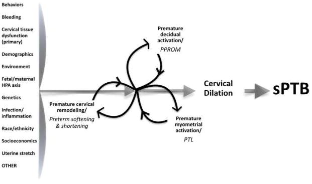
The cervix functions as a barrier between the uterus and vagina and keeps the uterus closed until term so that the fetus can develop. For delivery the cervix must soften and dilate, and finally reconstitute to close the uterus. This complex process involves precisely timed activation of molecular and microstructural events. Spontaneous preterm birth (sPTB) can result from aberrant timing of these events in the cervix. Unfortunately, the pathophysiology of sPTB due to cervical causes remains unclear and thus our treatment options remain limited - even if all appropriate candidates were identified and correctly treated with currently available interventions, the rate of sPTB would only be reduced by 5%. Very recent molecular and microstructural investigation is challenging prevailing concepts about cervical remodeling in pregnancy. We believe that progress toward novel, targeted solutions for the diverse pathways to sPTB entails a paradigm shift in which the overlapping and complex interactions between the cervix, uterus, membranes, fetus, placenta, and surrounding (structural and molecular) environment are suitably honored.
Keywords: Cervical insufficiency; Cervical microstructural assessment; Cervical remodeling; Cervical ripening; Spontaneous preterm birth.
Copyright © 2016 Elsevier Ltd. All rights reserved.
Conflict of interest statement
None declared.
Figures
References
-
- Danforth DN. The morphology of the human cervix. Clin Obstet Gynecol. 1983;26:7–13. - PubMed
-
- Dubrauszky V, Schwalm H, Fleischer M. Fibre system of connective tissue in childbearing age, menopause and pregnancy. Archiv Gynakol. 1971;210:276–92. - PubMed
-
- Aspden RM. Collagen organization in the cervix and its relation to mechanical function. Coll Relat Res. 1988;8:103–12. - PubMed
Publication types
MeSH terms
Grants and funding
LinkOut - more resources
Full Text Sources
Other Literature Sources
Medical
