

The Flagellum Attachment Zone: 'The Cellular Ruler' of Trypanosome Morphology
- PMID: 26776656
- PMCID: PMC4827413
- DOI: 10.1016/j.pt.2015.12.010
The Flagellum Attachment Zone: 'The Cellular Ruler' of Trypanosome Morphology
Abstract
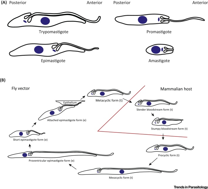
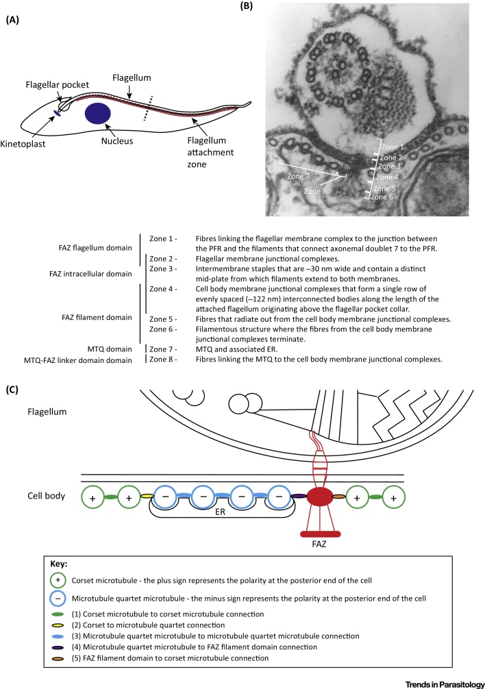
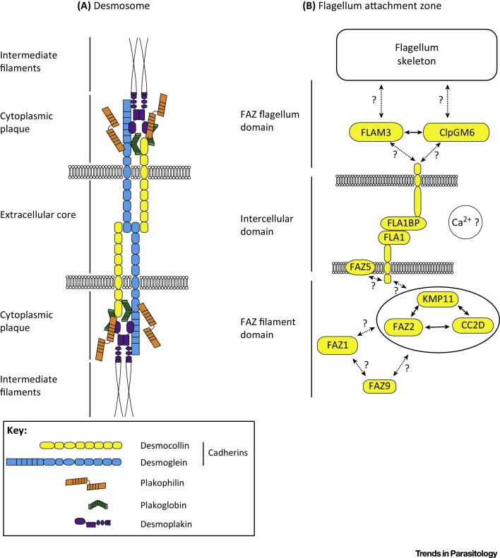
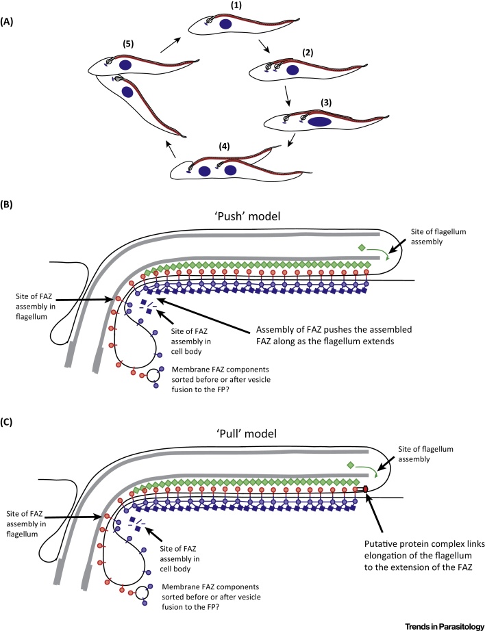
A defining feature of Trypanosoma brucei cell shape is the lateral attachment of the flagellum to the cell body, mediated by the flagellum attachment zone (FAZ). The FAZ is a complex cytoskeletal structure that connects the flagellum skeleton through two membranes to the cytoskeleton. The FAZ acts as a 'cellular ruler' of morphology by regulating cell length and organelle position and is therefore critical for both cell division and life cycle differentiations. Here we provide an overview of the advances in our understanding of the composition, assembly, and function of the FAZ.
Keywords: FAZ; Trypanosoma brucei; cytoskeleton; flagellum; morphology.
Copyright © 2016 The Authors. Published by Elsevier Ltd.. All rights reserved.
Figures
Similar articles
-
A dynamic coordination of flagellum and cytoplasmic cytoskeleton assembly specifies cell morphogenesis in trypanosomes.J Cell Sci. 2015 Apr 15;128(8):1580-94. doi: 10.1242/jcs.166447. Epub 2015 Mar 3. J Cell Sci. 2015. PMID: 25736289 Free PMC article.
-
An intracellular membrane junction consisting of flagellum adhesion glycoproteins links flagellum biogenesis to cell morphogenesis in Trypanosoma brucei.J Cell Sci. 2013 Jan 15;126(Pt 2):520-31. doi: 10.1242/jcs.113621. Epub 2012 Nov 23. J Cell Sci. 2013. PMID: 23178943
-
Assembly and maintenance of the flagellum attachment zone filament in Trypanosoma brucei.J Cell Sci. 2015 Jul 1;128(13):2361-72. doi: 10.1242/jcs.168377. Epub 2015 May 13. J Cell Sci. 2015. PMID: 25972344 Free PMC article.
-
Assembly of the flagellum and its role in cell morphogenesis in Trypanosoma brucei.Curr Opin Microbiol. 2010 Aug;13(4):453-8. doi: 10.1016/j.mib.2010.05.006. Epub 2010 Jun 10. Curr Opin Microbiol. 2010. PMID: 20541452 Review.
-
Cell-to-flagellum attachment and surface architecture in kinetoplastids.Trends Parasitol. 2023 May;39(5):332-344. doi: 10.1016/j.pt.2023.02.009. Epub 2023 Mar 16. Trends Parasitol. 2023. PMID: 36933967 Review.
Cited by
-
Novel Cytoskeleton-Associated Proteins in Trypanosoma brucei Are Essential for Cell Morphogenesis and Cytokinesis.Microorganisms. 2021 Oct 27;9(11):2234. doi: 10.3390/microorganisms9112234. Microorganisms. 2021. PMID: 34835360 Free PMC article.
-
The kinesin of the flagellum attachment zone in Leishmania is required for cell morphogenesis, cell division and virulence in the mammalian host.PLoS Pathog. 2021 Jun 18;17(6):e1009666. doi: 10.1371/journal.ppat.1009666. eCollection 2021 Jun. PLoS Pathog. 2021. PMID: 34143858 Free PMC article.
-
TbSmee1 regulates hook complex morphology and the rate of flagellar pocket uptake in Trypanosoma brucei.Mol Microbiol. 2018 Feb;107(3):344-362. doi: 10.1111/mmi.13885. Epub 2017 Dec 18. Mol Microbiol. 2018. PMID: 29178204 Free PMC article.
-
Characterisation of TbSmee1 suggests endocytosis allows surface-bound cargo to enter the trypanosome flagellar pocket.J Cell Sci. 2023 Oct 15;136(20):jcs261548. doi: 10.1242/jcs.261548. Epub 2023 Oct 26. J Cell Sci. 2023. PMID: 37737012 Free PMC article.
-
Use of chiral cell shape to ensure highly directional swimming in trypanosomes.PLoS Comput Biol. 2017 Jan 31;13(1):e1005353. doi: 10.1371/journal.pcbi.1005353. eCollection 2017 Jan. PLoS Comput Biol. 2017. PMID: 28141804 Free PMC article.
References
-
- Vickerman K. On the surface coat and flagellar adhesion in trypanosomes. J. Cell Sci. 1969;5:163–193. - PubMed
-
- Sherwin T., Gull K. The cell division cycle of Trypanosoma brucei brucei: timing of event markers and cytoskeletal modulations. Philos. Trans. R. Soc. Lond. B Biol. Sci. 1989;323:573–588. - PubMed
-
- Woods A. Definition of individual components within the cytoskeleton of Trypanosoma brucei by a library of monoclonal antibodies. J. Cell Sci. 1989;93:491–500. - PubMed
-
- Vaughan S. A repetitive protein essential for the flagellum attachment zone filament structure and function in Trypanosoma brucei. Protist. 2008;159:127–136. - PubMed
-
- Zhou Q. A coiled-coil- and C2-domain-containing protein is required for FAZ assembly and cell morphology in Trypanosoma brucei. J. Cell Sci. 2011;124:3848–3858. - PubMed
Publication types
MeSH terms
Grants and funding
LinkOut - more resources
Full Text Sources
Other Literature Sources
