. 2016;25(5):811-27.
doi: 10.3727/096368916X690467. Epub 2016 Jan 15.

Melanoma Immunotherapy in Mice Using Genetically Engineered Pluripotent Stem Cells

Affiliations

Melanoma Immunotherapy in Mice Using Genetically Engineered Pluripotent Stem Cells

Mohammad Haque et al. Cell Transplant. 2016.

Abstract

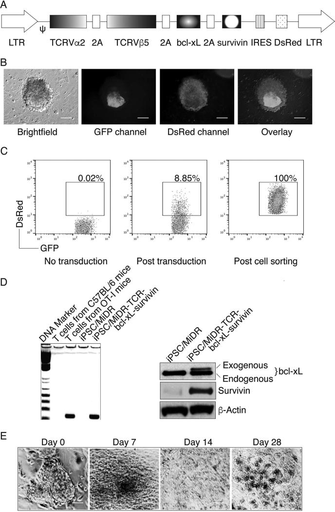
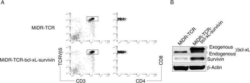
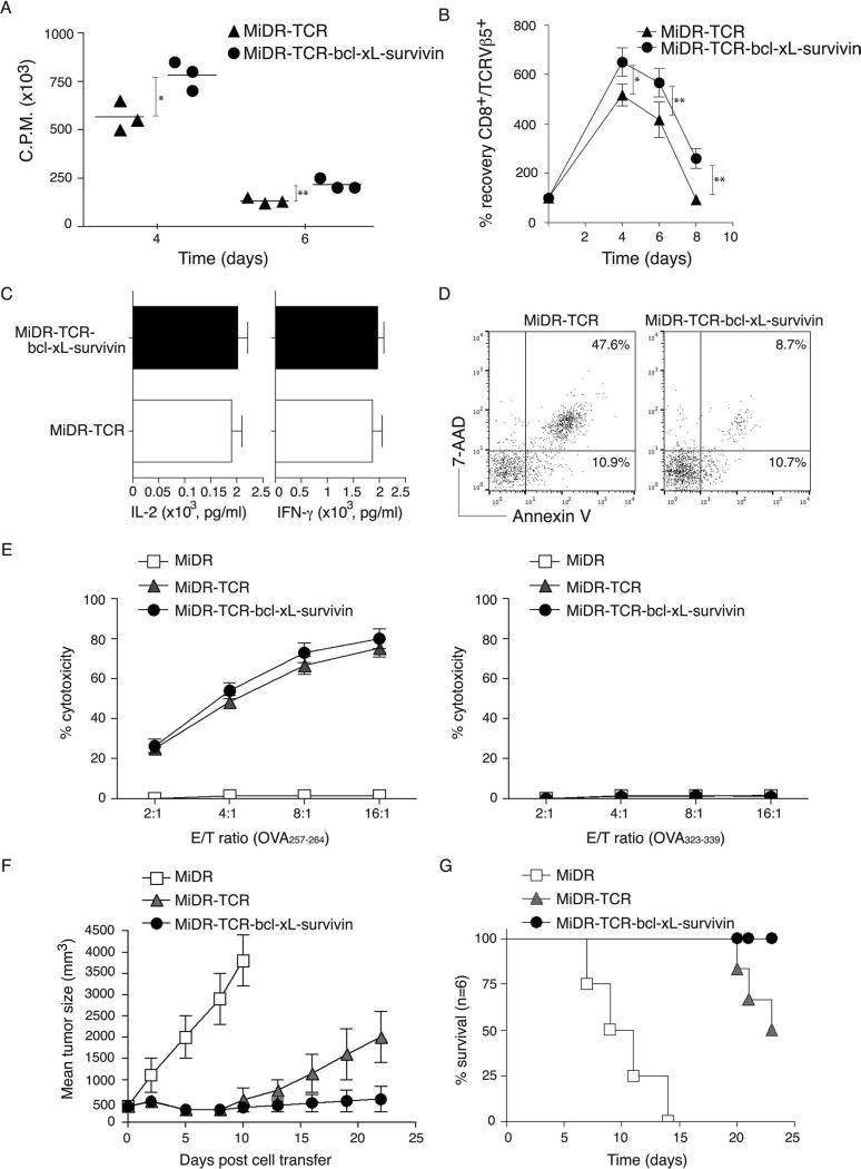
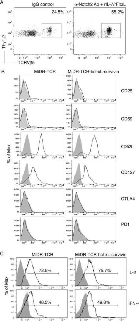
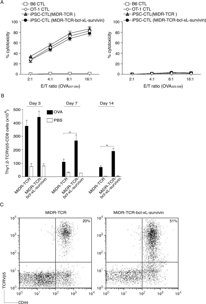
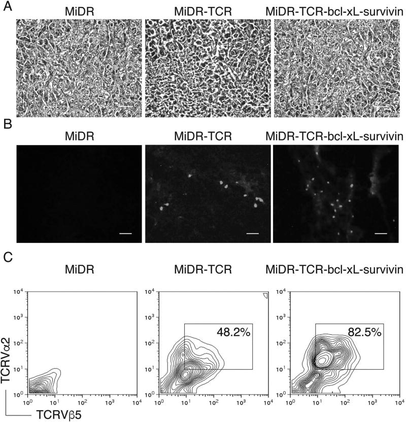
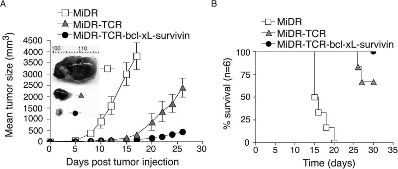
Adoptive cell transfer (ACT) of antigen (Ag)-specific CD8(+) cytotoxic T lymphocytes (CTLs) is a highly promising treatment for a variety of diseases. Naive or central memory T-cell-derived effector CTLs are optimal populations for ACT-based immunotherapy because these cells have a high proliferative potential, are less prone to apoptosis than terminally differentiated cells, and have the higher ability to respond to homeostatic cytokines. However, such ACT with T-cell persistence is often not feasible due to difficulties in obtaining sufficient cells from patients. Here we present that in vitro differentiated HSCs of engineered PSCs can develop in vivo into tumor Ag-specific naive CTLs, which efficiently suppress melanoma growth. Mouse-induced PSCs (iPSCs) were retrovirally transduced with a construct encoding chicken ovalbumin (OVA)-specific T-cell receptors (TCRs) and survival-related proteins (i.e., BCL-xL and survivin). The gene-transduced iPSCs were cultured on the delta-like ligand 1-expressing OP9 (OP9-DL1) murine stromal cells in the presence of murine recombinant cytokines (rFlt3L and rIL-7) for a week. These iPSC-derived cells were then intravenously adoptively transferred into recipient mice, followed by intraperitoneal injection with an agonist α-Notch 2 antibody and cytokines (rFlt3L and rIL-7). Two weeks later, naive OVA-specific CD8(+) T cells were observed in the mouse peripheral lymphatic system, which were responsive to OVA-specific stimulation. Moreover, the mice were resistant to the challenge of B16-OVA melanoma induction. These results indicate that genetically modified stem cells may be used for ACT-based immunotherapy or serve as potential vaccines.


Conflict of interest statement

The authors declare no conflicts of interest.

Figures

Figure 1
Figure 1
In vitro generation of highly reactive antigen-specific induced pluripotent stem cell-derived cytotoxic lymphocytes (Ag-specific iPSC-CTLs). (A) Schematic representation of the retroviral construct MiDR-TCRVα2-2A-TCRVβ5-2A-BCL-xL-2A-survivin expressing ovalbumin (OVA)-specific T-cell receptor (TCR), B-cell chronic lymphocytic leukemia (CLL)/lymphoma-extra large (BCL-xL), and survivin. Ψ, packaging signal; 2A, picornavirus self-cleaving 2A sequence; LTR, long terminal repeats. (B) The gene-transduced induced pluripotent stem cells (iPSCs) (GFP+DsRed+) were visualized by fluorescence microscopy (scale bars: 50 µm). Nanog promoter-driven green fluorescent protein (GFP) serves as a marker of the iPSCs. (C) GFP+DsRed+ iPSCs were transduced with the retroviral construct and GFP+DsRed+ iPSCs (middle) were analyzed by flow cytometry and sorted by a high-speed cell sorter (right). (D) GFP+DsRed+ iPSCs were sorted, and genomic DNA was analyzed for the TCRVβ5 gene by PCR (left). The expression of BCL-xL, survivin, and β-actin was determined by Western blotting. Data are representative of three independent experiments. (E) Morphology of T-cell differentiation on days 0, 7, 14, and 28 after cocultures of iPSCs transduced with genes of the TCR, BCL-xL, and survivin on the delta-like ligand 1-expressing OP9 murine stromal cells (OP9-DL1 cells) in the presence of murine recombinant Fms-like tyrosine kinase 3 ligand (rFlt3L) and recombinant interleukin-7 (rIL-7) (scale bars: 20 µm). Data are representative of three independent experiments.
Figure 2
Figure 2
Highly reactive Ag-specific iPSC-CTLs stably express BCL-xL and survivin. The iPSCs transduced with the MiDR-TCRVα2-2-TCRVβ5 (MiDR-TCR) or MiDR-TCRVα2-2A-TCRVβ5-2A-BCL-xL-2A-survivin (MiDR-TCR-BCL-xL-survivin) were cocultured with the OP9-DL1 cells in the presence of murine rFlt3L and rIL-7. On day 28 of in vitro coculture, the CD3+TCRβ5+CD8+ T cells were analyzed by flow cytometry and Western blotting. (A) Flow cytometric analysis of cluster of differentiation 3 (CD3) and TCRβ5, after gating on live iPSC-derived cells, and CD4 and CD8, after gating on CD3+TCRβ5+ populations. Data are representative of three independent experiments. (B) The expression of BCL-xL, survivin, and β-actin was determined by Western blotting. Data are representative of three independent experiments.
Figure 3
Figure 3
Functional analyses of highly reactive Ag-specific iPSC-CTLs. On day 28 of in vitro coculture, the single positive (SP) CD8+TCRβ5+ iPSC-CTLs were sorted and stimulated by T-depleted splenocytes pulsed with OVA257–264 peptide, and the proliferation, cytokine production, survival, and cytotoxicity were assessed. In some experiments, the sorted SP CD8+TCRβ5+ iPSC-CTLs were adoptively transferred into C57BL/6 mice that had been SC injected in the flank region with B16-OVA melanoma cells for a week and irradiated prior to the cell transfer. Mice were subsequently IV infected with recombinant vaccinia viruses expressing the gene for OVA (VV-OVA) and IP administered rIL-2 after the cell transfer. (A) Proliferation on day 1 to day 7 was measured by pulsing with tritiated thymidine for the last 20 h. Data are mean cpm ± SD from triplicate cultures and are representative of three experiments (*p < 0.05, **p < 0.01, Student’s unpaired t-test). (B) Survival based on recovery of DsRed+Vβ5+ T cells over time. Cell numbers present on day 0 were assigned a value of 100%, and cell numbers surviving on day 4 to day 8 were used to calculate the recovery percentage. The data represent the mean ± SD percentage change from three separate experiments (*p < 0.05, Student’s unpaired t-test). Data are representative of three independent experiments. (C) IL-2 and interferon (IFN)-γ production were measured by enzyme-linked immunosorbent assay (ELISA) at 40 h. Data are representative of three independent experiments. (D) Apoptosis of DsRed+CD8+ T cells on day 6 based on staining of annexin V and 7-aminoactinomycin D (7-AAD) and analyzed by flow cytometry. Data are representative of three independent experiments. (E) In vitro cytotoxicity assay. The iPSC-CTLs or control cells were added at different effector to target cell (E:T) ratios. Analysis was performed after a 12-h incubation period. Data are mean ± SD from three wells and representative of three experiments. (F) Tumor growth was monitored over time. Tumor volume was calculated as follows: V = length × width2 × 0.52. Data are mean tumor size ± SD from six individual mice and representative of three experiments. (G) Mouse survival was assessed over 25 days (Kaplan–Meier survival analysis).
Figure 4
Figure 4
In vivo development of highly reactive Ag-specific iPSC-CTLs. The iPSCs transduced with the MiDR-TCR or MiDR-TCR-BCL-xL-survivin were cocultured with the OP9-DL1 cells in the presence of murine rFlt3L and rIL-7. On day 7, the DsRed+ cells that were sorted were adoptively transferred into Thy1.1 congenic mice, and on the following days, mice were IP injected with agonistic α-Notch2 Ab, rIL-7, and rFlt3L or a mouse IgG/PBS control twice a week. (A) After 2 weeks, Thy1.2+TCRVβ5+ cells from the pooled lymph nodes and spleen were analyzed by flow cytometry, after gating on CD8+ T-cell population. A representative image from mice receiving the iPSCs transduced with the MiDR-TCR-BCL-xL-survivin is shown following IgG control or agonistic α-Notch2 Ab, rIL-7, and rFlt3L protein injections. (B) Expression of CD25, CD69, CD62L, CD127, CTL4, and PD1 was analyzed by flow cytometry, after gating on CD8+Thy1.2+TCRVβ5+ T cells from the pooled lymph nodes and spleen (dark lines; shaded areas indicate isotype controls). Data are representative of three independent experiments. (C) IL-2 and IFN-γ production (dark lines; shaded areas indicate isotype controls). The pooled lymph nodes and spleen were stimulated with OVA257–264 peptide and analyzed by intracellular cytokine staining, after gating on Thy1.2+TCRVβ5+ cells. Data are representative of three independent experiments.
Figure 5
Figure 5
Highly reactive Ag-specific iPSC-CTLs persist in vivo. In vivo development of highly reactive Ag-specific iPSC-CTLs was performed as described in Figure 4. Two weeks later, after the iPSC transfer and the in vivo Notch signaling, CD8+Thy1.2+TCRVβ5+ T cells from the pooled lymph nodes and spleen were sorted, and an in vitro cytotoxicity assay was performed. In some experiments, mice were subsequently IP challenged with whole OVA protein (100 µg) in PBS (filled bars) or with PBS alone (open bars). (A) In vitro cytotoxicity assay. The iPSC-CTLs, OVA257–264-specific CTLs from OT-I TCR Tg mice, or nonspecific CTLs from B6 mice were added at different effector to target cell (E:T) ratios. Analysis was performed after a 12-h incubation period. Data are mean ± SD from three wells and representative of three experiments. (B) On days 3, 7, and 14, Thy1.2+TCRVβ5+CD8+ T cells were counted from the pooled lymph nodes and spleen. Data are the mean number of Thy1.2+TCRVβ5+CD8+ cells ± SD from six individual mice and representative of three experiments (*p < 0.05, Student’s unpaired t-test). (C) At day 14, the percentage of TCRVβ5+CD44+ T cells was analyzed by flow cytometry, after gating on live CD8+ T cells in the spleen. Results are representative of three experiments.
Figure 6
Figure 6
Highly reactive Ag-specific iPSC-CTLs developed in vivo infiltrate into tumor tissues. In vivo development of highly reactive Ag-specific iPSC-CTLs was performed as described in Figure 4. Two weeks later, after the iPSC transfer and the in vivo Notch signaling, mice were SC injected in the flank region with B16-OVA melanoma cells that express the OVA Ag recognizable by the OVA257–264-specific CTLs. On day 21 to 22 after tumor challenge, tumor tissues were examined for tumor-reactive T-cell infiltration. (A) Hematoxylin and eosin (H&E) staining (scale bars: 20 µm). (B) Immunohistological staining (scale bars: 20 µm). OVA-specific TCRVα2+ CTLs (gray) infiltrated in OVA-expressing tumor tissues (the dark background). (C) Single-cell suspensions from tumor tissues were analyzed for expression of TCRVα2+ and TCRVβ5+ by flow cytometry, after gating on the CD8+ population. Data are representative of three independent experiments.
Figure 7
Figure 7
Highly reactive Ag-specific iPSC-CTLs developed in vivo suppress tumor growth. In vivo development of highly reactive Ag-specific iPSC-CTLs was performed as described in Figure 3. Two weeks later, after the iPSC transfer and the in vivo Notch signaling, mice were SC injected in the flank region with B16-OVA melanoma cells that express the OVA Ag recognizable by the OVA257–264-specific CTLs. Mice were subsequently IV infected with VV-OVA and IP administered rIL-2 after tumor inoculation twice per day for 3 days. (A) Tumor growth was monitored over time. Data are mean tumor size ± SD from six individual mice and representative of three experiments. Tumor representatives from the three groups at day 15 were shown. (B) Mouse survival was assessed over 30 days (Kaplan–Meier survival analysis).

Similar articles

Cited by

References

    1. Bargou R, Leo E, Zugmaier G, Klinger M, Goebeler M, Knop S, Noppeney R, Viardot A, Hess G, Schuler M, Einsele H, Brandl C, Wolf A, Kirchinger P, Klappers P, Schmidt M, Riethmuller G, Reinhardt C, Baeuerle PA, Kufer P. Tumor regression in cancer patients by very low doses of a T cell-engaging antibody. Science. 2008;321(5891):974–977. - PubMed
    1. Barrett DM, Singh N, Porter DL, Grupp SA, June CH. Chimeric antigen receptor therapy for cancer. Annu. Rev. Med. 2014;65:333–347. - PMC - PubMed
    1. Bartlett EK, Fetsch PA, Filie AC, Abati A, Steinberg SM, Wunderlich JR, White DE, Stephens DJ, Marincola FM, Rosenberg SA, Kammula US. Human melanoma metastases demonstrate nonstochastic site-specific antigen heterogeneity that correlates with T-cell infiltration. Clin. Cancer Res. 2014;20(10):2607–2616. - PMC - PubMed
    1. Beerman I, Bhattacharya D, Zandi S, Sigvardsson M, Weissman IL, Bryder D, Rossi DJ. Functionally distinct hematopoietic stem cells modulate hematopoietic lineage potential during aging by a mechanism of clonal expansion. Proc. Natl. Acad. Sci. USA. 2010;107(12):5465–5470. - PMC - PubMed
    1. Bendle GM, Linnemann C, Hooijkaas AI, Bies L, de Witte MA, Jorritsma A, Kaiser AD, Pouw N, Debets R, Kieback E, Uckert W, Song JY, Haanen JB, Schumacher TN. Lethal graft-versus-host disease in mouse models of T cell receptor gene therapy. Nat. Med. 2010;16(5):565–570. - PubMed

Publication types