. 2016 Jan 18:6:19371.
doi: 10.1038/srep19371.

Isolation of highly enriched primary human microglia for functional studies

Affiliations

Isolation of highly enriched primary human microglia for functional studies

Justin Rustenhoven et al. Sci Rep. .

Abstract

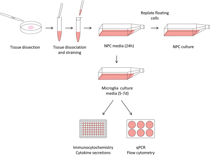
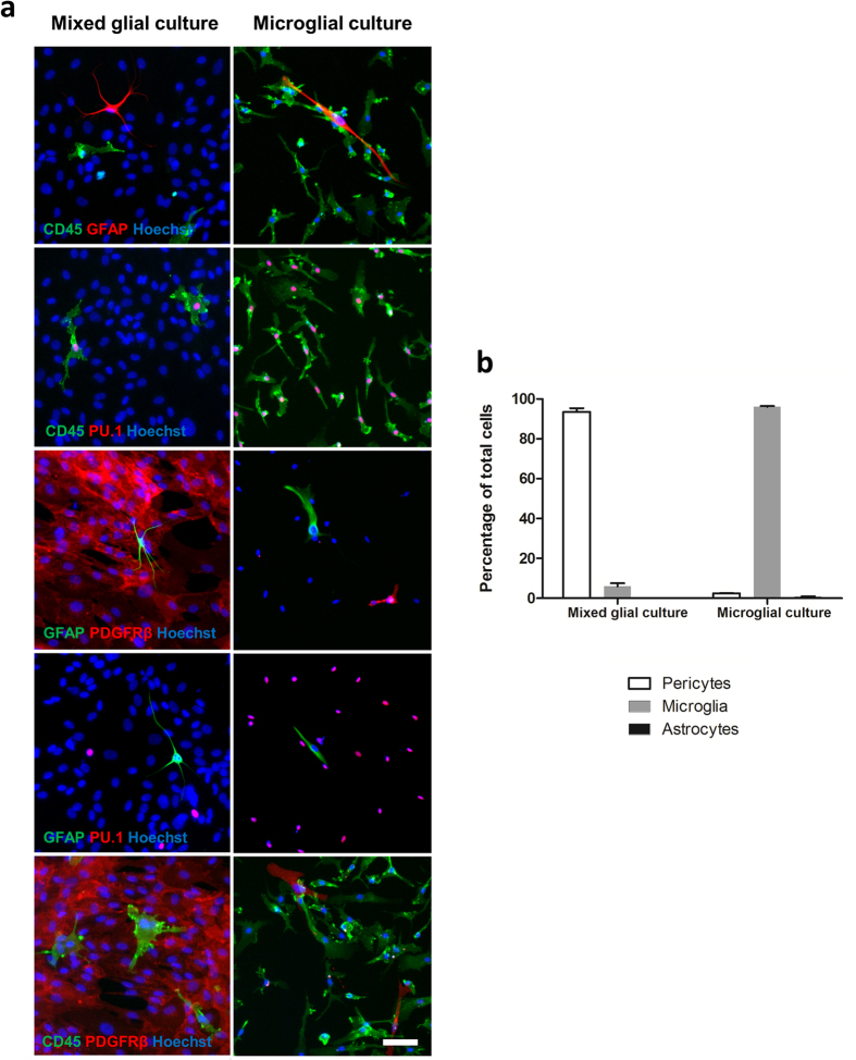
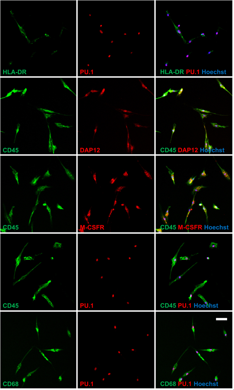
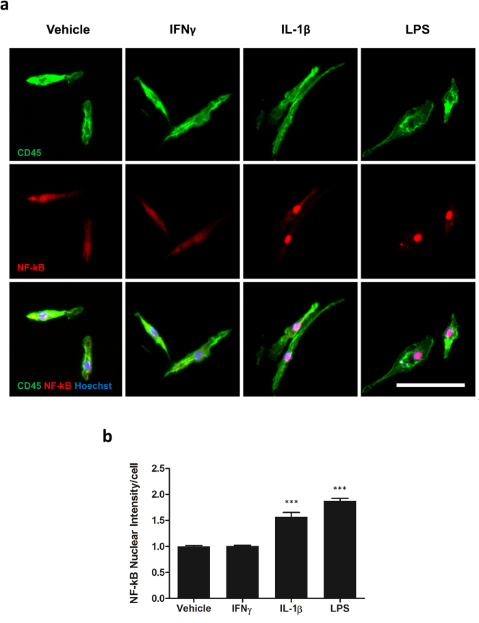
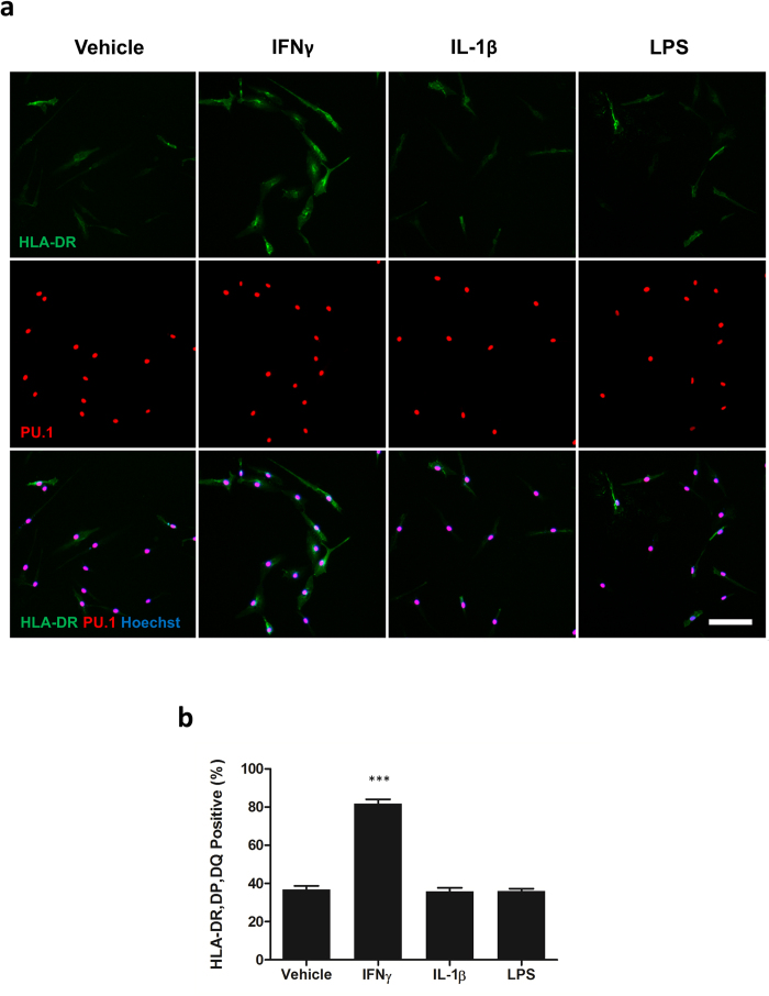
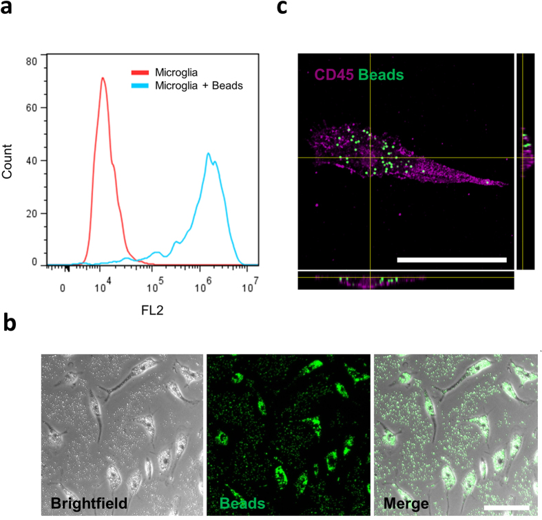
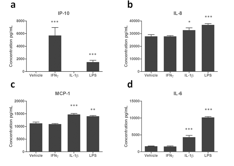
Microglia, the resident macrophages of the central nervous system play vital roles in brain homeostasis through clearance of pathogenic material. Microglia are also implicated in neurological disorders through uncontrolled activation and inflammatory responses. To date, the vast majority of microglial studies have been performed using rodent models. Human microglia differ from rodent counterparts in several aspects including their response to pharmacological substances and their inflammatory secretions. Such differences highlight the need for studies on primary adult human brain microglia and methods to isolate them are therefore required. Our procedure generates microglial cultures of >95% purity from both biopsy and autopsy human brain tissue using a very simple media-based culture procedure that takes advantage of the adherent properties of these cells. Microglia obtained in this manner can be utilised for research within a week. Isolated microglia demonstrate phagocytic ability and respond to inflammatory stimuli and their purity makes them suitable for numerous other forms of in vitro studies, including secretome and transcriptome analysis. Furthermore, this protocol allows for the simultaneous isolation of neural precursor cells during the microglial isolation procedure. As human brain tissue is such a precious and valuable resource the simultaneous isolation of multiple cell types is highly beneficial.


Figures

Figure 1
Figure 1. Overview of isolation procedure.
Human brain biopsy or autopsy tissue was mechanically and enzymatically dissociated to achieve a single cell suspension. This suspension was strained and plated in neural precursor cell media for up to 24 hours. Floating and weakly attached cells were removed and replated into a new flask and used to generate neural precursor cell cultures. Microglial culture media was added to the original flask for 5–7 days and microglia were harvested and utilised for experimental procedures.
Figure 2
Figure 2. Characterisation of culture purity.
Culture purity of our standard mixed glial isolation and microglial specific isolation was determined via immunocytochemistry using markers specific for microglia (CD45, PU.1), pericytes (PDGFRβ) and astrocytes (GFAP) (a). The percentages of each cell type were determined (b). N = 3 for mixed glial culture isolation (all epilepsy), N = 7 (three epilepsy, three GBM and one Parkinson’s disease) for microglia isolation. Scale bar = 50 μm.
Figure 3
Figure 3. Isolated microglia express typical microglial markers.
Isolated microglia were found to express markers present on microglia derived from mixed glial cultures. All CD45 or PU.1 positive cells co-expressed CD68, M-CSFR, DAP12 whilst displaying variable levels of HLA-DR. Representative images from three (all epilepsy) independent cases are shown. Scale bar = 50 μm.
Figure 4
Figure 4. Isolated microglia express cytoplasmic NF-κB under basal conditions which can be translocated to the nucleus by IL-1β and LPS but not IFNγ.
Microglia were treated with 10 ng/mL IL-1β, LPS, IFNγ or vehicle (0.1% BSA in PBS) for one hour and immunostained for NF-kB and CD45 (a). The intensity of nuclear NF-kB was determined (b). N = 3 (all epilepsy). Scale bar = 50 μm. ***p < 0.001.
Figure 5
Figure 5. Microglial HLA-DR, DP, DQ expression is induced by IFNγ but not IL-1β or LPS.
Microglia were treated with 10 ng/mL IL-1β, LPS, IFNγ or vehicle (0.1% BSA in PBS) for 24 hours and immunostained for HLA-DR, DP, DQ and PU.1 (a). The percentage of HLA, DR, DP, DQ positive cells was determined (b). N = 3 (two epilepsy and one GBM). Scale bar = 50 μm. ***p < 0.001.
Figure 6
Figure 6. Isolated microglia can phagocytose fluorescent beads.
Microglia were cultured in the presence or absence of fluorescent beads (1 μm diameter) for two hours. Cells were washed thoroughly to remove un-internalized beads and collected by trypsinisation and gentle scraping. Phagocytosis of beads was determined by a rightward shift in FL2 intensity via flow cytometry (a). One representative plot from three (two epilepsy and one GBM, frontal cortex) independent experiments is shown. Brightfield and fluorescent imaging of cells immediately prior to trypsinisation shows the distribution of beads within cells (b). Confocal scanning laser microscopy confirmed that microglia were able to internalise beads (c). Scale bar = 50 μm.
Figure 7
Figure 7. Measurement of cytokine/chemokine secretion from isolated microglia.
Microglia were treated with 10 ng/mL IL-1β, IFNγ, LPS or vehicle (0.1% BSA in PBS) for 24 hours. Conditioned media was collected and concentrations of IP-10 (a), IL-8 (b), MCP-1 (c) and IL-6 (d) determined in triplicate wells by a multiplex cytometric bead array. One representative case from three (all epilepsy) independent experiments is shown. *p < 0.05, **p < 0.01, ***p < 0.001.

References

    1. Ferguson T. A. & Griffith T. S. A vision of cell death: insights into immune privilege. Immunological reviews 156, 167–184 (1997). - PubMed
    1. del Rio-Hortega P. In Cytology and Cellular Pathology of the Nervous System (ed Penfield W.) Ch. 482–534, (Hoeber, 1932).
    1. Rezaie P. & Male D. Colonisation of the developing human brain and spinal cord by microglia: a review. Microscopy research and technique 45, 359–382, 10.1002/(sici)1097-0029(19990615)45:6<359::aid-jemt4>3.0.co;2-d(1999 ). - DOI - PubMed
    1. Yang I., Han S. J., Kaur G., Crane C. & Parsa A. T. The role of microglia in central nervous system immunity and glioma immunology. Journal of clinical neuroscience : official journal of the Neurosurgical Society of Australasia 17, 6–10, 10.1016/j.jocn.2009.05.006 (2010). - DOI - PMC - PubMed
    1. Kreutzberg G. W. Microglia: a sensor for pathological events in the CNS. Trends Neurosci 19, 312–318, 0166-2236(96)10049-7 (1996). - PubMed

Publication types