Review
. 2016:569:287-308.
doi: 10.1016/bs.mie.2015.07.015. Epub 2015 Aug 20.

In Vitro Model of the Epidermis: Connecting Protein Function to 3D Structure

Affiliations
Review

In Vitro Model of the Epidermis: Connecting Protein Function to 3D Structure

Christopher Arnette et al. Methods Enzymol. 2016.

Abstract

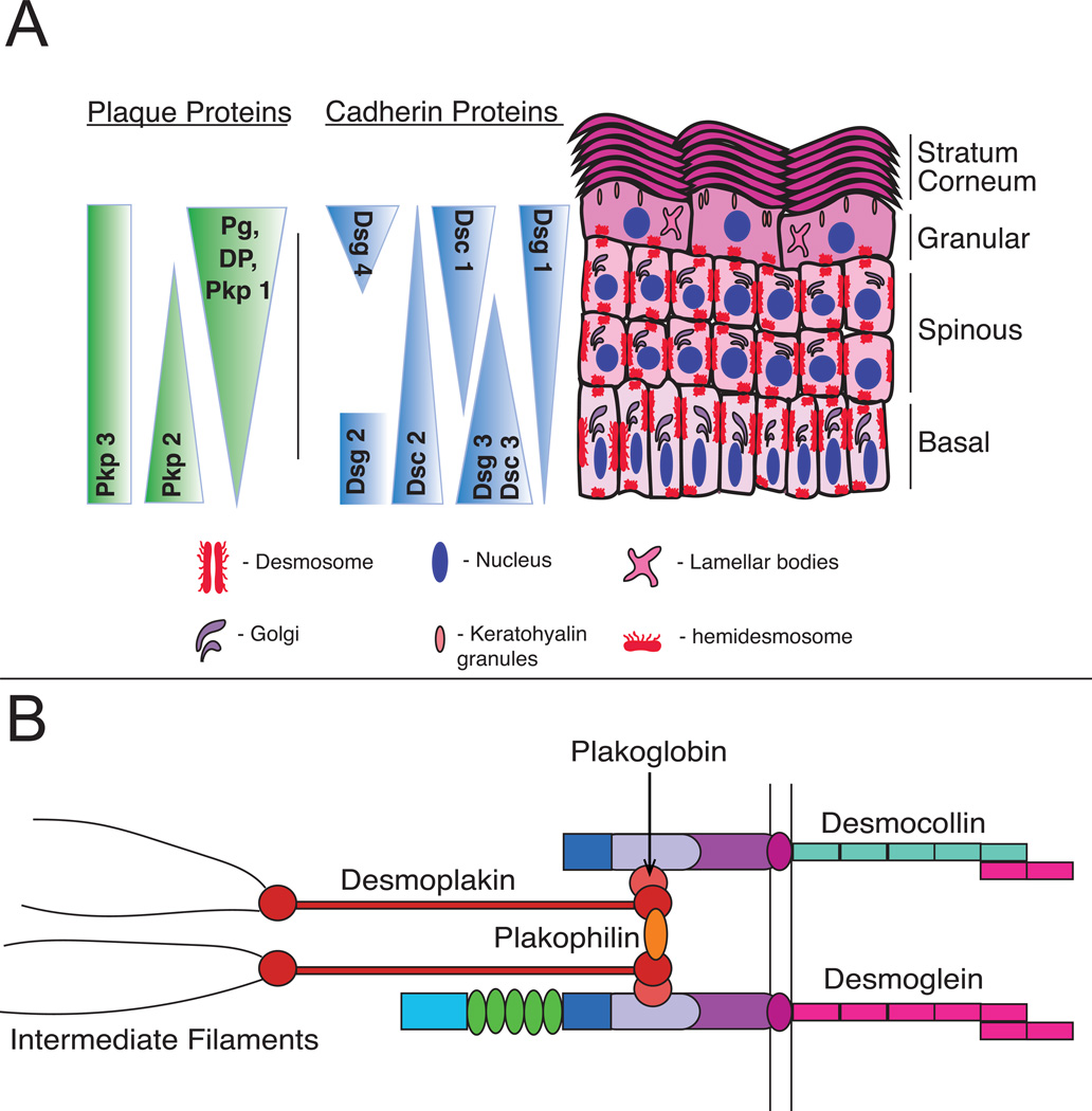
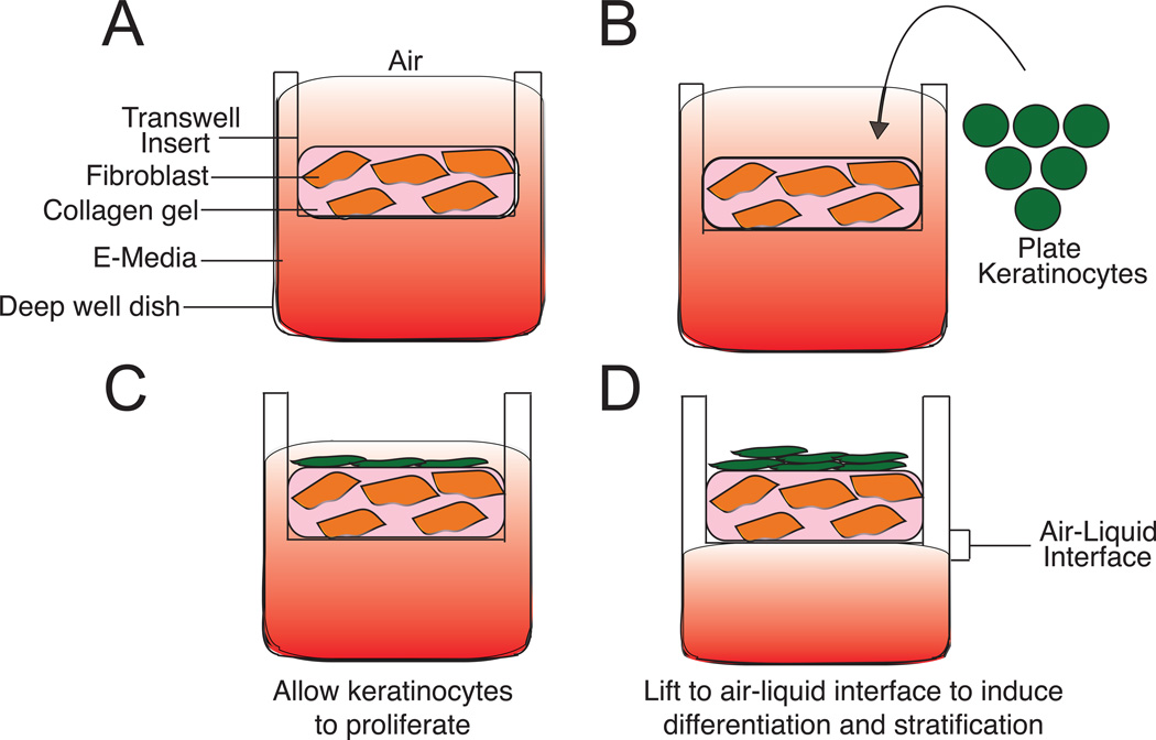
Much of our understanding of the biological processes that underlie cellular functions in humans, such as cell-cell communication, intracellular signaling, and transcriptional and posttranscriptional control of gene expression, has been acquired from studying cells in a two-dimensional (2D) tissue culture environment. However, it has become increasingly evident that the 2D environment does not support certain cell functions. The need for more physiologically relevant models prompted the development of three-dimensional (3D) cultures of epithelial, endothelial, and neuronal tissues (Shamir & Ewald, 2014). These models afford investigators with powerful tools to study the contribution of spatial organization, often in the context of relevant extracellular matrix and stromal components, to cellular and tissue homeostasis in normal and disease states.

Keywords: Desmoglein; Epidermis; Keratinocyte; Organotypic.


Figures

Figure 1
Figure 1. Organization and expression of desmosomal components within the skin
A) The are four layers within the stratified epidermis: basal, spinous, granular, and cornified. Each layer contains a complement of differentiation-specific junctional proteins critical to support epidermal cytoarchitecture and drive tissue morphogenesis. B) Desmosomes are specialized, calcium-dependent adhesive structures that are composed of three family of proteins: desmosomal cadherins, armadillo proteins, and plakin proteins. These structures link to the intermediate filament cytoskeleton to maintain tissue integrity.
Figure 2
Figure 2. Comparison to human epidermis and time course of 3D raft development
A) Cellular architecture of human skin and 3D organotypic raft culture. B) Keratinocytes were seeded on collagen plugs and were grown submerged conditions for 2 days. The cultures were then lifted to the air-liquid interface. Organotypic rafts were harvested 3, 6, 9, or 12 days after being lifted to air. Representative images of keratinocyte stratification in 3D raft cultures (Provided by Paul Hoover of the Northwestern University Skin Disease Research Core).
Figure 3
Figure 3. Generation of 3D organotypic cultures using the transwell method
A) A collagen-fibroblast plug is made in the upper chamber (transwell). Media is supplied from the top and bottom. B and C) Keratinocytes are seeded onto the collagen-fibroblast plug and kept under submerged conditions: media is supplied from the top and bottom chambers. Keratinocytes are allowed to proliferate. D) Organotypic rafts are raised to air-liquid interface. Media is only supplied from the bottom chamber.
Figure 4
Figure 4. Manipulation and analysis of Dsg1 in 3D organotypic raft cultures
A) H&E-stained sections of 6-day-old rafts expressing either miR Lmn or miR DG1. Silencing of DG1 results in altered suprabasal morphology (insets with continuous lines) and impaired differentiation (insets with dashed lines) compared to control. B) Western blot analysis of desmosomal proteins, desmoglein 1 and 3, lamin A/C (Lmn A/C), and GAPDH (glyceraldehyde-3-phosphate dehydrogenase) in 6-day-old 3D raft cultures. Either Dsg1 or Lamin expression was silenced in raft cultures using retroviruses engineered to express microRNA (miRNA)-like sequences specific for each protein. KD reveals ~90% reduction of protein levels under each condition. C) Real-time PCR analysis show decreased levels of desmocollin 1 mRNA levels following Dsg1 knockdown in 3D raft cultures, while a gene target of EGFR, EphA2, mRNA levels are increased. Note: Figure originally published in Getsios et al (© 2009 Getsios et al. Journal of Cell Biology. 185:1243-1258. doi:10.1083/jcb.200809044); Used with permission.

References

    1. Ader M, Tanaka EM. Modeling human development in 3D culture. Curr Opin Cell Biol. 2014;31:23–28. - PubMed
    1. Asselineau D, Prunieras M. Reconstruction of 'simplified' skin: control of fabrication. Br J Dermatol. 1984;111(Suppl 27):219–222. - PubMed
    1. Barreca A, De Luca M, Del Monte P, Bondanza S, Damonte G, Cariola G, Di Marco E, Giordano G, Cancedda R, Minuto F. In vitro paracrine regulation of human keratinocyte growth by fibroblast-derived insulin-like growth factors. J Cell Physiol. 1992;151(2):262–268. - PubMed
    1. Bilousova G, Roop DR. Generation of functional multipotent keratinocytes from mouse induced pluripotent stem cells. Methods Mol Biol. 2013;961:337–350. - PubMed
    1. Brooke MA, Etheridge SL, Kaplan N, Simpson C, O'Toole EA, Ishida-Yamamoto A, Marches O, Getsios S, Kelsell DP. iRHOM2-dependent regulation of ADAM17 in cutaneous disease and epidermal barrier function. Hum Mol Genet. 2014;23(15):4064–4076. - PMC - PubMed

Publication types

LinkOut - more resources