

Treatment Planning for Image-Guided Neuro-Vascular Interventions Using Patient-Specific 3D Printed Phantoms
- PMID: 26778878
- PMCID: PMC4712717
- DOI: 10.1117/12.2081997
Treatment Planning for Image-Guided Neuro-Vascular Interventions Using Patient-Specific 3D Printed Phantoms
Abstract
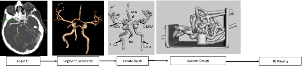
Minimally invasive endovascular image-guided interventions (EIGIs) are the preferred procedures for treatment of a wide range of vascular disorders. Despite benefits including reduced trauma and recovery time, EIGIs have their own challenges. Remote catheter actuation and challenging anatomical morphology may lead to erroneous endovascular device selections, delays or even complications such as vessel injury. EIGI planning using 3D phantoms would allow interventionists to become familiarized with the patient vessel anatomy by first performing the planned treatment on a phantom under standard operating protocols. In this study the optimal workflow to obtain such phantoms from 3D data for interventionist to practice on prior to an actual procedure was investigated. Patient-specific phantoms and phantoms presenting a wide range of challenging geometries were created. Computed Tomographic Angiography (CTA) data was uploaded into a Vitrea 3D station which allows segmentation and resulting stereo-lithographic files to be exported. The files were uploaded using processing software where preloaded vessel structures were included to create a closed-flow vasculature having structural support. The final file was printed, cleaned, connected to a flow loop and placed in an angiographic room for EIGI practice. Various Circle of Willis and cardiac arterial geometries were used. The phantoms were tested for ischemic stroke treatment, distal catheter navigation, aneurysm stenting and cardiac imaging under angiographic guidance. This method should allow for adjustments to treatment plans to be made before the patient is actually in the procedure room and enabling reduced risk of peri-operative complications or delays.
Keywords: 3D Printing; Additive Manufacturing; DSA; Image Guided Interventions; Neuro-vascular; Patient-Specific Phantoms; Treatment Planning; Vascular Phantoms.
Figures


Similar articles
-
3D Printed Abdominal Aortic Aneurysm Phantom for Image Guided Surgical Planning with a Patient Specific Fenestrated Endovascular Graft System.Proc SPIE Int Soc Opt Eng. 2017 Feb 11;10138:101380P. doi: 10.1117/12.2253902. Epub 2017 Mar 13. Proc SPIE Int Soc Opt Eng. 2017. PMID: 28638171 Free PMC article.
-
Design Optimization for Accurate Flow Simulations in 3D Printed Vascular Phantoms Derived from Computed Tomography Angiography.Proc SPIE Int Soc Opt Eng. 2017 Feb 11;10138:101380R. doi: 10.1117/12.2253711. Epub 2017 Mar 13. Proc SPIE Int Soc Opt Eng. 2017. PMID: 28663663 Free PMC article.
-
Challenges and limitations of patient-specific vascular phantom fabrication using 3D Polyjet printing.Proc SPIE Int Soc Opt Eng. 2014 Mar 13;9038:90380M. doi: 10.1117/12.2042266. Proc SPIE Int Soc Opt Eng. 2014. PMID: 25300886 Free PMC article.
-
3D-printed patient-specific applications in orthopedics.Orthop Res Rev. 2016 Oct 14;8:57-66. doi: 10.2147/ORR.S99614. eCollection 2016. Orthop Res Rev. 2016. PMID: 30774470 Free PMC article. Review.
-
[Diagnostic imaging of cerebrovascular disease on multi-detector row computed tomography (MDCT)].Brain Nerve. 2011 Sep;63(9):923-32. Brain Nerve. 2011. PMID: 21878694 Review. Japanese.
Cited by
-
2D vessel contrast dilution gradient (CDG) analysis using 1000 fps high speed angiography (HSA) for velocity distribution estimation.Proc SPIE Int Soc Opt Eng. 2022 Feb-Mar;12031:1203107. doi: 10.1117/12.2611790. Epub 2022 Apr 4. Proc SPIE Int Soc Opt Eng. 2022. PMID: 35982769 Free PMC article.
-
Hybrid computed tomography and magnetic resonance imaging 3D printed models for neurosurgery planning.Ann Transl Med. 2019 Nov;7(22):684. doi: 10.21037/atm.2019.10.109. Ann Transl Med. 2019. PMID: 31930085 Free PMC article. Review.
-
A simulation platform using 3D printed neurovascular phantoms for clinical utility evaluation of new imaging technologies.Proc SPIE Int Soc Opt Eng. 2018 Feb;10578:105780N. doi: 10.1117/12.2293630. Epub 2018 Mar 12. Proc SPIE Int Soc Opt Eng. 2018. PMID: 29887667 Free PMC article.
-
Biomimicking Atherosclerotic Vessels: A Relevant and (Yet) Sub-Explored Topic.Biomimetics (Basel). 2024 Feb 23;9(3):135. doi: 10.3390/biomimetics9030135. Biomimetics (Basel). 2024. PMID: 38534820 Free PMC article. Review.
-
Advanced 3D Mesh Manipulation in Stereolithographic Files and Post-Print Processing for the Manufacturing of Patient-Specific Vascular Flow Phantoms.Proc SPIE Int Soc Opt Eng. 2016 Feb 27;9789:978909. doi: 10.1117/12.2217036. Epub 2016 Mar 25. Proc SPIE Int Soc Opt Eng. 2016. PMID: 28649165 Free PMC article.
References
-
- Rudin S, Bednarek DR, Hoffmann KR. Endovascular image-guided interventions (EIGIs) Med Phys. 2008;35(1):301–309. http://www.ncbi.nlm.nih.gov/pubmed/18293585. - PMC - PubMed
-
- Deng J, Zhao Z, Gao G. Periprocedural complications associated with endovascular embolisation of intracranial ruptured aneurysms with matrix coils. Singapore Med J. 2007;48(5):429–433. - PubMed
-
- Nuis RJ, Piazza N, Van Mieghem NM, et al. In-hospital complications after transcatheter aortic valve implantation revisited according to the Valve Academic Research Consortium definitions. Catheter Cardiovasc Interv. 2011;78(3):457–467. - PubMed
-
- Guinot PG, Depoix JP, Etchegoyen L, et al. Anesthesia and perioperative management of patients undergoing transcatheter aortic valve implantation: analysis of 90 consecutive patients with focus on perioperative complications. J Cardiothorac Vasc Anesth. 2010;24(5):752–761. - PubMed
Grants and funding
LinkOut - more resources
Full Text Sources
Other Literature Sources