Review
. 2015 Dec 23:9:480.
doi: 10.3389/fncel.2015.00480. eCollection 2015.

Understanding How the Subcommissural Organ and Other Periventricular Secretory Structures Contribute via the Cerebrospinal Fluid to Neurogenesis

Affiliations
Review

Understanding How the Subcommissural Organ and Other Periventricular Secretory Structures Contribute via the Cerebrospinal Fluid to Neurogenesis

Maria M Guerra et al. Front Cell Neurosci. .

Abstract

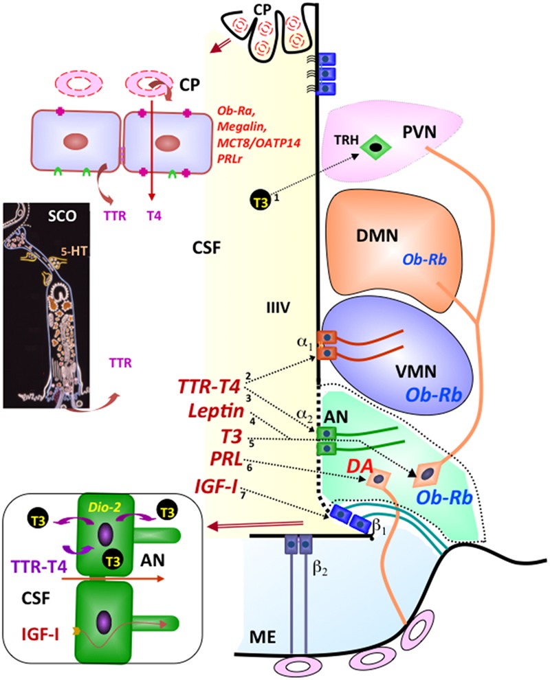
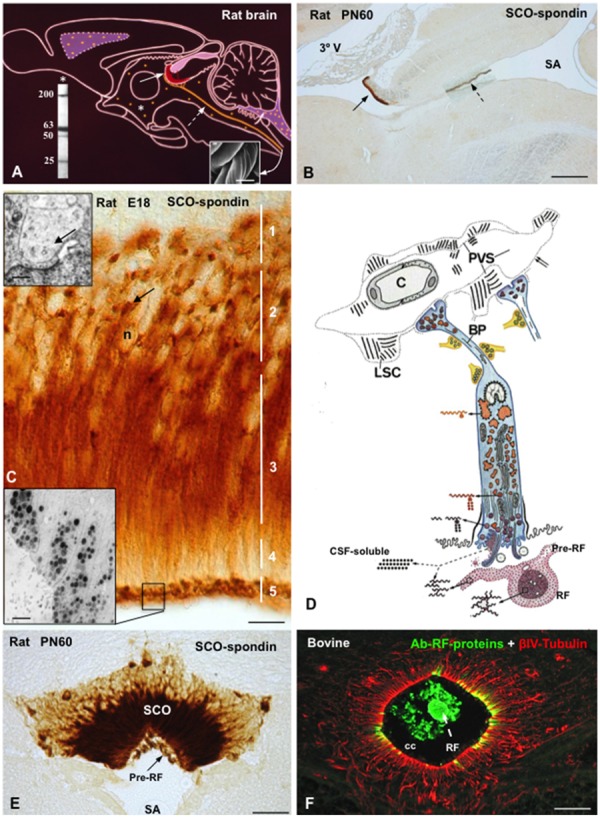
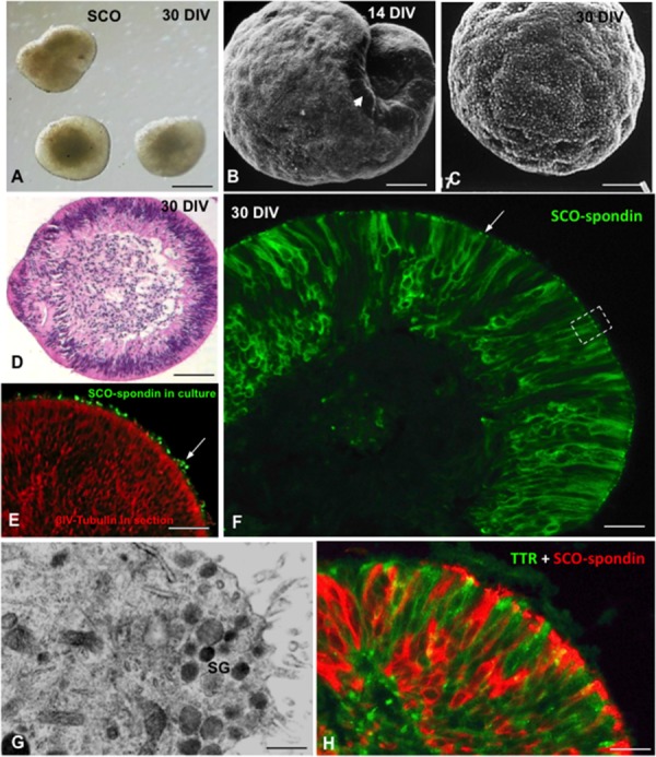
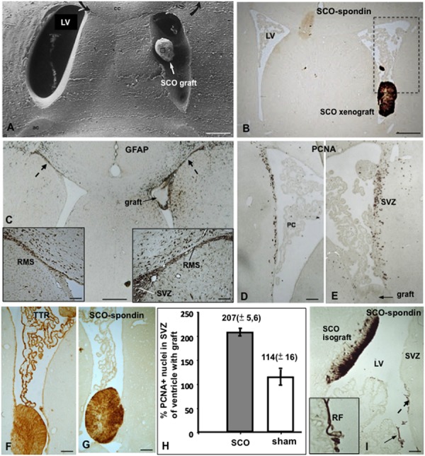
The dynamic and molecular composition of the cerebrospinal fluid (CSF) and, consequently, the CSF physiology is much more complex and fascinating than the simplistic view held for decades. Signal molecules either transported from blood to CSF or secreted into the CSF by circumventricular organs and CSF-contacting neurons, use the CSF to reach their targets in the brain, including the pre- and postnatal neurogenic niche. The subcommissural organ (SCO), a highly conserved brain gland present throughout the vertebrate phylum, is one of the sources for signals, as well as the choroid plexus, tanycytes and CSF-contacting neurons. The SCO secretes into the fetal and adult CSF SCO-spondin, transthyretin, and basic fibroblast growth factor. These proteins participate in certain aspects of neurogenesis, such as cell cycle of neural stem cells, neuronal differentiation, and axon pathfinding. Through the CSF, the SCO-secretory proteins may reach virtually any target in the embryonic and adult central nervous system. Since the SCO continues to secrete throughout life span, it seems likely that the neurogenetic property of the SCO compounds would be targeted to the niches where neurogenesis continues in adulthood. This review is aimed to bring into discussion early and new evidence concerning the role(s) of the SCO, and the probable mechanisms by which SCO compounds can readily reach the neurogenic niche of the subventricular zone flowing with the CSF to participate in the regulation of the neurogenic niche. As we unfold the multiples trans-fluid talks between discrete brain domains we will have more tools to influence such talks.

Keywords: CSF-contacting neurons; SCO-spondin; cerebrospinal fluid; circumventricular organs; integrins; neurogenesis; subcommissural organ; transthyretin.


Figures

FIGURE 1
FIGURE 1
Integrative pathways involving the CSF. By receptor mediated transport at the choroid plexus (CP), leptin (Ob-Ra), insulin growth factor I (megalin), thyroid hormones (MCT8/OATP14), and prolactin (PRLr) are transported from blood to CSF. Transthyretin (TTR) is secreted by choroid plexus and the subcommissural organ (SCO) into the CSF. The secretory activity of the SCO is under serotonin (5-HT) inhibitory control. Most CSF T4 is bound to TTR. TTR-T4 complexes are taken up by tanycytes that express deiodinase 2 (arrows 2, 3). Here (bottom left panel), T4 is converted to T3 and then released into the intercellular space of the arcuate nucleus (arrow 5) or into the CSF to reach the TRH-parvocellular neurons of the paraventricular nucleus (arrow 1). The milieu of the arcuate nucleus (AN; green background) is especially exposed to molecules present in the CSF and closed to the median eminence (ME) and ventromedial nucleus (VMN). Leptin present in the CSF may readily reach the neurons expressing the Ob-Rh receptor of the arcuate (arrow 4), ventromedial and dorsomedial nuclei of the hypothalamus. CSF prolactin (arrow 6) may reach the dopamine-secreting neurons (DA) of the arcuate nucleus that project to the portal capillaries of the median eminence (light-blue background). CSF insulin growth factor I (arrow 7) is internalized by β tanycytes and transported along their processes. Modified after Rodríguez et al. (2010).
FIGURE 2
FIGURE 2
The subcommissural organ is the phylogenetically oldest brain gland and the first to differentiate in ontogeny. (A–E) From amphioxus to primates, 500 million years of evolution. (A–C) Sagittal sections through the CNS of the amphioxus (Branchiostoma lanceolatum, Acrania), showing the location (A) and immunoreactivity (B,C) of the cells forming the Infundibular organ (IO). V, ventricle; cc, central canal; from Olsson et al. (1994). (C) Line drawing of the CNS of the amphioxus showing a secretory ependyma in the recessus neuroporicus (a), the infundibular organ (b) and the central canal with Reissner fiber (c); from Olsson and Wingstrand (1954). (D) Subcommissural organ and Reissner fiber (arrow) of the primate Aotes. SA, sylvius aqueduct; from Rodríguez et al. (1993). (E) Sagittal section through the epithalamus of a 13-weeks-old human fetus immunostained with an antiserum against a 45 kDa compound (most likely corresponding to TTR) obtained from the CSF of a hydrocephalic fetus. A population of ependymocytes are strongly immunoreactive; from Rodríguez et al. (1993). Right inset detailed magnification of previous figure showing immunoreactive (arrow) and immunonegative (asterisk) ependymal cells; left inset SCO from a 32 GW fetus immunostained for SCO-spondin; all cells are immunoreactive (arrow). (F) Sagittal section through the CNS of a Xenopus l larvae. The cells of the subcommissural organ (SCO) and the floor plate (FP) strongly express SCO-spondin. (G) Sagittal section through the CNS of a 3-days-old chick embryo. A small group of neuroependymal cells located a the roof of the diencephalic vesicle (Di) expresses SCO-spondin (arrow). Te, telencephalon; Mc, mesencephalon. (H) Detailed view of previous figure showing that SCO-spondin is mainly located in the apical region of the neuroependymal cells (arrow). (i) At the 7th day of incubation, the chick SCO is fully differentiated with SCO-spondin located in the cell body of ependymocytes (broken arrow) and along their basal processes ending at the pial membrane (full arrows). PC, posterior commissure; from Schoebitz et al. (1986). Scale bars: (A,B) 80 μm; (C) 16 μm; (D) 400 μm; (E) 100 μm; right Inset 9 μm; left inset 8 μm; (F) 300 μm; (G) 280 μm; (H) 56 μm; (I) 85 μm.
FIGURE 3
FIGURE 3
The subcommissural organ-Reissner fiber complex. (A) Drawing depicting the rat subcommissural organ (red, full arrow)- Reissner fiber (orange, broken arrow) complex and the CSF-soluble secretion (orange dots, asterisk). (Right inset) Scanning electron microscopy of bovine RF collected from the central canal. Left inset. Western blot of CSF of PN30 rats, immunoreacted with antibodies against SCO-spondin. CSF-soluble compounds of 200, 63, 50, and 25 kDa of molecular weight are shown. (B) Sagittal section of a rat brain immunostained with anti-SCO-spondin at postnatal day 60. The SCO (full arrow)-RF (broken arrow) complex is selectively immunoreactive. (C) High magnification of the SCO of a rat embryo (E18) immunostained with anti-SCO-spondin. Zonation of a SCO-cell (1–5) is shown. Arrow points to paranuclear immunoreactive masses corresponding to RER. Upper inset. Electron microscopy of dilated RER cisternae (arrow). Lower inset. Electron microscopy immunocytochemistry using anti-SCO spondin showing secretory granules stored at the apical cell pole. (D) Drawing depicting the ultrastructure and the secretory process of a SCO-ependymal cell. They are bipolar cells, with and apical pole contacting the ventricular CSF and a basal process projecting to local capillaries and to the subarachnoid space. Glycoproteins secreted by the SCO cells either remain soluble into CSF or polymerize forming the RF. The secretory material upon release condenses, first as a film on the surface of the organ (pre-RF) and, after further packaging, into RF. The basal processes of ependymal cells (BP) receive abundant serotonergic, gabaergic, and catecholaminergic neural inputs and end on a network of basal lamina containing long spacing collagen (LSC). PVS, perivascular space. (E) Frontal section of a rat brain at PN60 immunostained with anti-SCO-spondin. SCO and pre-RF are strongly reactive. (F) Frontal section of the bovine spinal cord processed for double immunofluorescence using anti-RF proteins (green) and βIV-tubulin (red). The central canal (cc) contains Reissner fiber (RF, green) and is lined by tanycytes-like ependymal cells (red). Scale bars: (B) 200 μm; (C) 10 μm; (E) 40 nm; (F) 20 μm. From Rodríguez et al. (1993); Vío et al. (2008), Ortloff et al. (2013).
FIGURE 4
FIGURE 4
The cerebrospinal fluid is a pathway for the delivery of neurotropic factors to the adult SVZ niche. (A) Cell organization of SVZ niche in the adult brain. SVZ astrocytes (B, blue) are stem cells which generate migrating neuroblasts (A, red) destined for the olfactory bulb via rapidly dividing transit-amplifying cells (C, green). A specialized basal lamina (BL, black) extends from perivascular cells and contacts all cell types, including multiciliated ependyma cells (E, orange). Ependymal cells, neural terminals (ne), the extracellular matrix (ECM)-basal lamina (BL) network, and the cerebrospinal fluid (CSF) are key components of the niche and regulator of the adult neurogenesis. Stem cells display a single 9+0 cilium to sensor CSF signals. Compounds secreted into the CSF by circumventricular organs such as the subcommissural organ (SCO) and choroid plexus (CP), or by CSF-contacting neurons can readily reach the SVZ (modified after Riquelme et al., 2008). (B) Frontal section of the rat SCO immunostained with antibodies against SCO-spondin and βIV-tubulin (from Ortloff et al., 2013). (C) Choroid plexus immunostained for TTR. (D) Drawing depicting a hypothalamic peptidergic CSF-contacting neuron with a dendrite projecting to the ventricle bearing a 9+0 cilium, and axon projecting to the capillaries of the pituitary gland and bearing and axonal branch reaching the ventricle (from Rodríguez, 1976). (E) Electron microscopy of a peptide terminal within the ventricle, with neurosecretory granules undergoing exocytosis. Scale bars: (B) 120 μm; (C) 35 μm; (E) 700 nm.
FIGURE 5
FIGURE 5
Multidomain organization of thrombosponin type 1 molecules. LDL receptor domains are indicated by the yellow box. EGF like domains are indicated by the red box. Thrombospondin types 1, 2, and 3 repeats (TSRs) are indicated by the blue boxes. A number of cellular and extracellular binding molecules for the domains have been identified. Many of these are components of ECM. CBM, cellular binding molecules; EBM, extracellular binding molecules.
FIGURE 6
FIGURE 6
(A) Ligands for integrin-β1 heterodimers. Many of these ligands are components of ECM. (B) Simplified schematic drawing of how SCO-spondin might promote neurogenesis in the adult SZV niche. SCO-spondin (1) may change the composition of ECM (i.e., transforming the type of collagen) and (2) the availability of growth factors in the niche, modifying (3) the immediate microenvironment and behavior of niche cells. Some of these functions could be mediated (4) by interaction of SCO-spondin with integrin-β1 signaling and (5) cross talking with other essential pathways, like those regulated by bFGF and TTR/thyroid hormones (6).
FIGURE 7
FIGURE 7
Organ culture of bovine subcommissural organ. After 30 days in culture, SCO explants organize forming spheres of secretory ependymocytes. (A) Phase contrast microscopy. (B,C) Scanning electron microscopy after 14 (B) and 30 DIV (C). (D) Section of a SCO-explant stained with haematoxylin-eosin. (E) Secretory evidence of secretion. Explants were cultured in the presence of antibodies against SCO-spondin. After histological procedure, sections were incubated with anti-IgG conjugated with alexa 488. Immunofluorescence reveals the presence of SCO-spondin aggregates associated to cilia (green, arrow). (F) Section of a SCO-explant immunostained for SCO-spondin showing the intracellular and extracellular (arrow) location of the protein. (G) Ultrathin section of an area similar to that framed in previous figure, showing the ultrastructure of the apical cell pole loaded with secretory granules (sg). (H) Section of a SCO-explant. Double immunofluorescence for SCO-spondin (red) and TTR (green). Scale bars: (A) 60 μm; (B–E) 25 μm; (F) 10 μm; (G) 500 nm; (H) 10 μm. From Schöebitz et al. (2001), Montecinos et al. (2005).
FIGURE 8
FIGURE 8
Xeno- and isografting of SCO-explants into the lateral ventricle of adult rats. (A–H) Bovine SCO-explants 30 DIV were grafted into the lateral ventricle of adult rats. (A) Scanning electron microscopy showing a SCO-explant in the ventricle. (B) Frontal section of the brain of a grafted animal immunostained with AFRU. The grafted SCO is strongly reactive. The area framed is shown in figures D and E. LV, lateral ventricle. (C) Frontal section of the brain of a grafted animal immunostained for GFAP. Astrocytes forming the rostral migratory stream (RMS) are shown. In the grafted ventricle the RMS is hypertrophied (right inset) as compared to that of the contralateral ventricle (left inside). (D,E) Areas similar to that framed in figure (B), immunostained for PCNA. In the grafted ventricle (E) proliferation is significantly higher than in the contralateral ventricle (D). (F,G) The grafted SCO expresses TTR and SCO-spondin. TTR is also expressed by the choroid plexus. (H) Quantitative analysis of PCNA+ nuclei after SCO grafting in a lateral ventricle of an adult normal rat. The results are expressed as percentage of the number of labeled nuclei in the SVZ of the ventricle carrying the grafts with respect to that of the contralateral ventricle, taken as 100%. Sham operated rats underwent surgery as for transplantation, but received no graft. There is a twofold increase of PCNA+ nuclei in the grafted ventricle. (I) Rat SCO explant grafted into the lateral ventricle of an adult rat. The graft becomes integrated into the wall of the lateral ventricle (LV) with the ependymal cells secreting SCO-spondin into the ventricle aggregated on the ependyma of the subventricular zone (broken arrow; SVZ) and forming a Reissner fiber (RF; full arrow; inset). Scale bars: (A–C) 120 μm; (D,E) 60 μm; (F,G) 40 μm; (I) 60 μm. From Rodríguez et al. (1999); González (2007).

Similar articles

Cited by

References

    1. Abrous D. N., Koehl M., Le Moal M. (2005). Adult neurogenesis: from precursors to network and physiology. Physiol. Rev. 85 523–569. 10.1152/physrev.00055.2003 - DOI - PubMed
    1. Adams J. C. (2001). Thrombospondins: multifunctional regulators of cell interaction. Annu. Rev. Cell. Dev. Biol. 17 25–51. 10.1146/annurev.cellbio.17.1.25 - DOI - PubMed
    1. Almazan G., Honegger P., Matthieu J. M. (1985). Triiodothyronine stimulation of oligodendroglial differentiation and myelination. A developmental study. Dev. Neurosci. 7 45–54. 10.1159/000112275 - DOI - PubMed
    1. Alshehri B., D’Souza D. G., Lee J. Y., Petratos S., Richardson S. J. (2015). The diversity of mechanisms influenced by transthyretin in neurobiology: development, disease and endocrine disruption. J. Neuroendocrinol. 27 303–323. 10.1111/jne.12271 - DOI - PubMed
    1. Alvarez-Buylla A., Garcia-Verdugo J. M. (2002). Neurogenesis in adult subventricular zone. J. Neurosci. 22 629–634. - PMC - PubMed