Neonatal Perirhinal Lesions in Rhesus Macaques Alter Performance on Working Memory Tasks with High Proactive Interference
- PMID: 26778978
- PMCID: PMC4700260
- DOI: 10.3389/fnsys.2015.00179
Neonatal Perirhinal Lesions in Rhesus Macaques Alter Performance on Working Memory Tasks with High Proactive Interference
Abstract
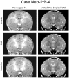
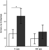
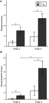
The lateral prefrontal cortex is known for its contribution to working memory (WM) processes in both humans and animals. Yet, recent studies indicate that the prefrontal cortex is part of a broader network of interconnected brain areas involved in WM. Within the medial temporal lobe (MTL) structures, the perirhinal cortex, which has extensive direct interactions with the lateral and orbital prefrontal cortex, is required to form active/flexible representations of familiar objects. However, its participation in WM processes has not be fully explored. The goal of this study was to assess the effects of neonatal perirhinal lesions on maintenance and monitoring WM processes. As adults, animals with neonatal perirhinal lesions and their matched controls were tested in three object-based (non-spatial) WM tasks that tapped different WM processing domains, e.g., maintenance only (Session-unique Delayed-nonmatching-to Sample, SU-DNMS), and maintenance and monitoring (Object-Self-Order, OBJ-SO; Serial Order Memory Task, SOMT). Neonatal perirhinal lesions transiently impaired the acquisition of SU-DNMS at a short (5 s) delay, but not when re-tested with a longer delay (30 s). The same neonatal lesions severely impacted acquisition of OBJ-SO task, and the impairment was characterized by a sharp increase in perseverative errors. By contrast, neonatal perirhinal lesion spared the ability to monitor the temporal order of items in WM as measured by the SOMT. Contrary to the SU-DNMS and OBJ-SO, which re-use the same stimuli across trials and thus produce proactive interference, the SOMT uses novel objects on each trial and is devoid of interference. Therefore, the impairment of monkeys with neonatal perirhinal lesions on SU-DNMS and OBJ-SO tasks is likely to be caused by an inability to solve working memory tasks with high proactive interference. The sparing of performance on the SOMT demonstrates that neonatal perirhinal lesions do not alter working memory processes per se but rather impact processes modulating impulse control and/or behavioral flexibility.
Keywords: excitotoxic lesion; perseveration; proactive interference; self-ordered task; serial order memory.
Figures


Similar articles
-
Impaired Cognitive Flexibility After Neonatal Perirhinal Lesions in Rhesus Macaques.Front Syst Neurosci. 2019 Jan 30;13:6. doi: 10.3389/fnsys.2019.00006. eCollection 2019. Front Syst Neurosci. 2019. PMID: 30760985 Free PMC article.
-
Neonatal hippocampal lesions in rhesus macaques alter the monitoring, but not maintenance, of information in working memory.Behav Neurosci. 2011 Dec;125(6):859-70. doi: 10.1037/a0025541. Epub 2011 Sep 19. Behav Neurosci. 2011. PMID: 21928873 Free PMC article.
-
Object and spatial memory after neonatal perirhinal lesions in monkeys.Behav Brain Res. 2016 Feb 1;298(Pt B):210-7. doi: 10.1016/j.bbr.2015.11.010. Epub 2015 Nov 23. Behav Brain Res. 2016. PMID: 26593109 Free PMC article.
-
Development and plasticity of the neural circuitry underlying visual recognition memory.Can J Physiol Pharmacol. 1995 Sep;73(9):1364-71. doi: 10.1139/y95-191. Can J Physiol Pharmacol. 1995. PMID: 8748986 Review.
-
The perirhinal cortex and long-term familiarity memory.Q J Exp Psychol B. 2005 Jul-Oct;58(3-4):234-45. doi: 10.1080/02724990444000122. Q J Exp Psychol B. 2005. PMID: 16194967 Review.
Cited by
-
Persistent negative effects of alcohol drinking on aspects of novelty-directed behavior in male rhesus macaques.Alcohol. 2017 Sep;63:19-26. doi: 10.1016/j.alcohol.2017.03.002. Epub 2017 Jun 23. Alcohol. 2017. PMID: 28847378 Free PMC article.
-
Proactive interference and the development of working memory.Wiley Interdiscip Rev Cogn Sci. 2022 May;13(3):e1593. doi: 10.1002/wcs.1593. Epub 2022 Feb 22. Wiley Interdiscip Rev Cogn Sci. 2022. PMID: 35193170 Free PMC article. Review.
-
Reevaluating the role of the hippocampus in memory: A meta-analysis of neurotoxic lesion studies in nonhuman primates.Hippocampus. 2023 Jun;33(6):787-807. doi: 10.1002/hipo.23499. Epub 2023 Jan 17. Hippocampus. 2023. PMID: 36649170 Free PMC article.
-
Off to a Good Start: The Early Development of the Neural Substrates Underlying Visual Working Memory.Front Syst Neurosci. 2016 Aug 18;10:68. doi: 10.3389/fnsys.2016.00068. eCollection 2016. Front Syst Neurosci. 2016. PMID: 27587999 Free PMC article. Review.
-
Neonatal perirhinal cortex lesions impair monkeys' ability to modulate their emotional responses.Behav Neurosci. 2017 Oct;131(5):359-71. doi: 10.1037/bne0000208. Behav Neurosci. 2017. PMID: 28956946 Free PMC article.
References
-
- Bertolino A., Saunders R., Mattay V., Bachevalier J., Frank J., Weinberger D. (1997). Altered development of prefrontal neurons in rhesus monkeys with neonatal mesial temporo-limbic lesions: a proton magnetic resonance spectroscopic imaging study. Cereb. Cortex 7, 740–748. 10.1093/cercor/7.8.740 - DOI - PubMed
Grants and funding
LinkOut - more resources
Full Text Sources
Other Literature Sources
Research Materials

