Review
. 2016 Jan 5:9:181.
doi: 10.3389/fnsys.2015.00181. eCollection 2015.

Role of Prefrontal Persistent Activity in Working Memory

Affiliations
Review

Role of Prefrontal Persistent Activity in Working Memory

Mitchell R Riley et al. Front Syst Neurosci. .

Abstract

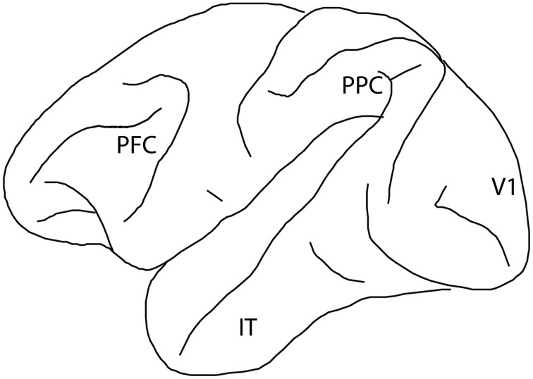
The prefrontal cortex is activated during working memory, as evidenced by fMRI results in human studies and neurophysiological recordings in animal models. Persistent activity during the delay period of working memory tasks, after the offset of stimuli that subjects are required to remember, has traditionally been thought of as the neural correlate of working memory. In the last few years several findings have cast doubt on the role of this activity. By some accounts, activity in other brain areas, such as the primary visual and posterior parietal cortex, is a better predictor of information maintained in visual working memory and working memory performance; dynamic patterns of activity may convey information without requiring persistent activity at all; and prefrontal neurons may be ill-suited to represent non-spatial information about the features and identity of remembered stimuli. Alternative interpretations about the role of the prefrontal cortex have thus been suggested, such as that it provides a top-down control of information represented in other brain areas, rather than maintaining a working memory trace itself. Here we review evidence for and against the role of prefrontal persistent activity, with a focus on visual neurophysiology. We show that persistent activity predicts behavioral parameters precisely in working memory tasks. We illustrate that prefrontal cortex represents features of stimuli other than their spatial location, and that this information is largely absent from early cortical areas during working memory. We examine memory models not dependent on persistent activity, and conclude that each of those models could mediate only a limited range of memory-dependent behaviors. We review activity decoded from brain areas other than the prefrontal cortex during working memory and demonstrate that these areas alone cannot mediate working memory maintenance, particularly in the presence of distractors. We finally discuss the discrepancy between BOLD activation and spiking activity findings, and point out that fMRI methods do not currently have the spatial resolution necessary to decode information within the prefrontal cortex, which is likely organized at the micrometer scale. Therefore, we make the case that prefrontal persistent activity is both necessary and sufficient for the maintenance of information in working memory.

Keywords: fMRI; monkey; neuron; neurophysiology; prefrontal cortex.


Figures

Figure 1
Figure 1
Diagram of the monkey brain, with four cortical regions implicated in visual working memory labeled: prefrontal cortex (PFC), posterior parietal cortex (PPC), primary visual cortex (V1), and inferior temporal cortex (IT).
Figure 2
Figure 2
Schematic diagram of intrinsic connections between neurons within the prefrontal cortex. Neurons with similar tuning (memory field representing upper right location) are drawn in red color. Pyramidal neurons excite each other through reciprocal connections. Stripes of neurons with similar spatial tuning are repeated across the surface of the cortex. Interneurons inhibit other pyramidal neurons with different spatial tuning (memory field representing lower right location) drawn in blue color.
Figure 3
Figure 3
(A) Sequence of events in the Oculomotor Delayed Response (ODR) task. Successive frames represent the fixation period, stimulus presentation, delay period, and saccade toward the remembered stimulus location. (B) Delayed Match to Sample task. Monkeys first foveate the fixation point and pull a lever. They are then presented with a cue stimulus. This is followed by a random (0–2) number of non-match stimuli, separated by delay periods. When a match stimulus appears at the same location as the cue, the monkeys are required to release the lever. (C) Match/Non-match task. While monkeys fixate, two stimuli are presented in sequence, separated by delay periods. After another delay period, two choice targets are shown and the monkey has to saccade to the green target if the second stimulus matched the cue, and the blue stimulus, otherwise. (D) Schematic diagram of prefrontal activity elicited by the stimulus that is sustained during the delay period in each of the previous tasks.
Figure 4
Figure 4
(A) Simulated, network activity in the ODR task, following presentation of a cue at the 180° location. Abscissa represents time during the trial; ordinate represents different neurons arranged based on their tuning. (B) Network activity illustrating drifts in the peak of activation during the delay period. Axes have been rotated relative to (A). Color represents firing rate. The black triangle represents the cue position at the beginning of the delay period (encoded population activity on the bottom graph). The red triangle represents the location decoded by the population activity at the end of the delay period. (C) Left, saccade endpoints in one behavioral session divided into trials that landed clockwise (red) or counterclockwise (blue) relative to the cue stimulus position. Right, delay-period responses of one neuron recorded during the same session. The triangles indicate the circular mean of the tuning curve obtained from trials that generated clockwise, or counterclockwise saccadic deviations. (D) Left, schematic representation of four different delay period population activity profiles to the same 180° cue. Red lines represent trials with saccadic endpoints closer to the target (accurate trials) and green lines represent trials farther from the target (inaccurate trials). Right, difference between discharge variability in inaccurate and accurate trials depending on the location of the cue. Variability is maximal for cue appearing at the flanks of the neuron's tuning curve, where small deviations cause large differences in firing rate. (E) Left, schematic representation of delay period activity of two neurons recorded simultaneously, whose tuning peaks lie at opposite sides of the activity bump. Right, trial-to-trial correlations are negative between these neurons as a bump in activity leads to an increase in firing rate of one neuron with a decrease in the other neuron. Panel (A) adapted with permission from Renart et al. (2003); panels (B–E) from Wimmer et al. (2014).

Similar articles

Cited by

References

    1. Albers A. M., Kok P., Toni I., Dijkerman H. C., De Lange F. P. (2013). Shared representations for working memory and mental imagery in early visual cortex. Curr. Biol. 23, 1427–1431. 10.1016/j.cub.2013.05.065 - DOI - PubMed
    1. Arnsten A. F., Cai J. X., Goldman-Rakic P. S. (1988). The alpha-2 adrenergic agonist guanfacine improves memory in aged monkeys without sedative or hypotensive side effects: evidence for alpha-2 receptor subtypes. J. Neurosci. 8, 4287–4298. - PMC - PubMed
    1. Arnsten A. F., Goldman-Rakic P. S. (1985). Alpha 2-adrenergic mechanisms in prefrontal cortex associated with cognitive decline in aged nonhuman primates. Science 230, 1273–1276. 10.1126/science.2999977 - DOI - PubMed
    1. Arnsten A. F., Wang M. (2016). Targeting prefrontal cortical systems for drug development: potential therapies for cognitive disorders. Annu. Rev. Pharmacol. Toxicol. [Epub ahead of print]. - PMC - PubMed
    1. Atkinson R. C., Shiffrin R. M. (1968). Human memory: a proposed system and its control processes, in The Psychology of Learning and Motivation, eds Spence K. W., Spence J. T. (London: Academic Press; ), 89–195.

LinkOut - more resources