. 2016 Jan 5:6:1479.
doi: 10.3389/fmicb.2015.01479. eCollection 2015.

Probing an Interfacial Surface in the Cyanide Dihydratase from Bacillus pumilus, A Spiral Forming Nitrilase

Affiliations

Probing an Interfacial Surface in the Cyanide Dihydratase from Bacillus pumilus, A Spiral Forming Nitrilase

Jason M Park et al. Front Microbiol. .

Abstract

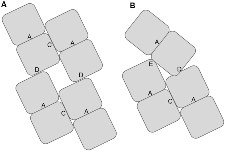
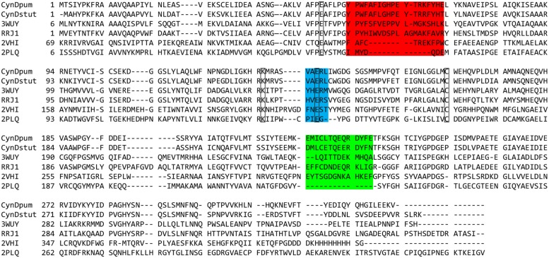
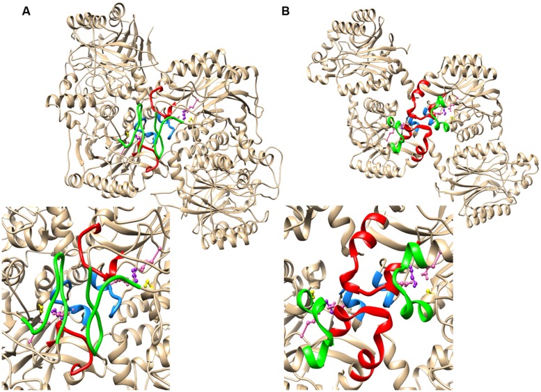
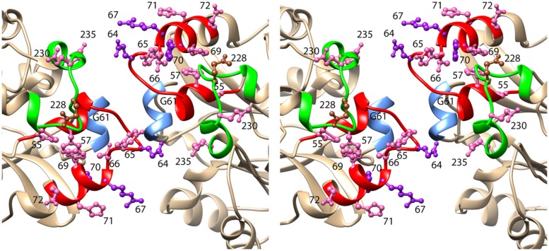
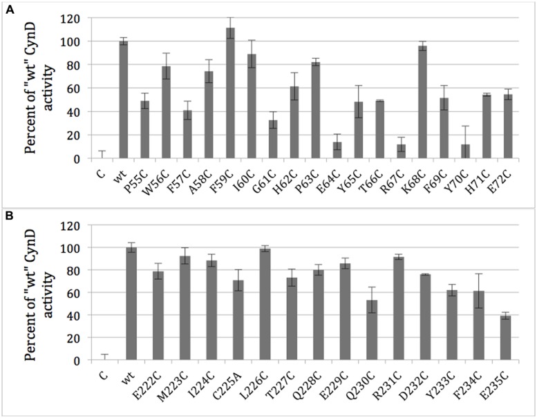
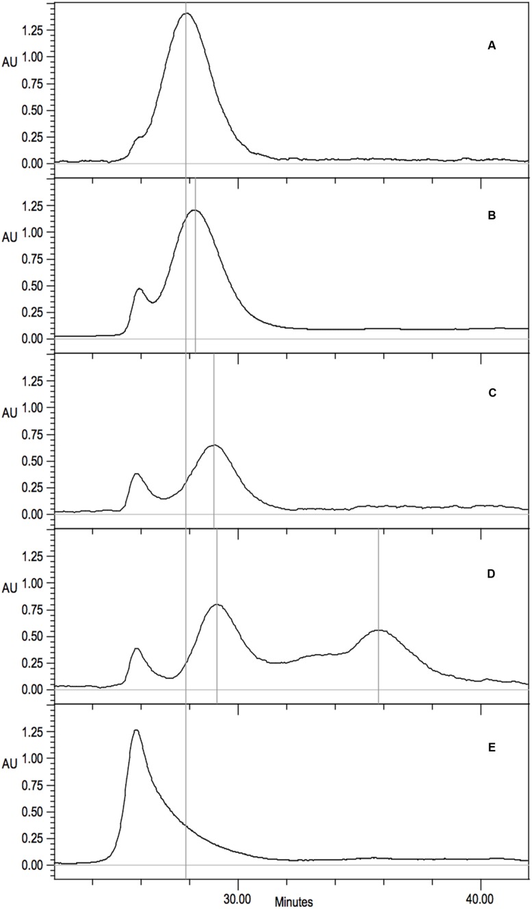
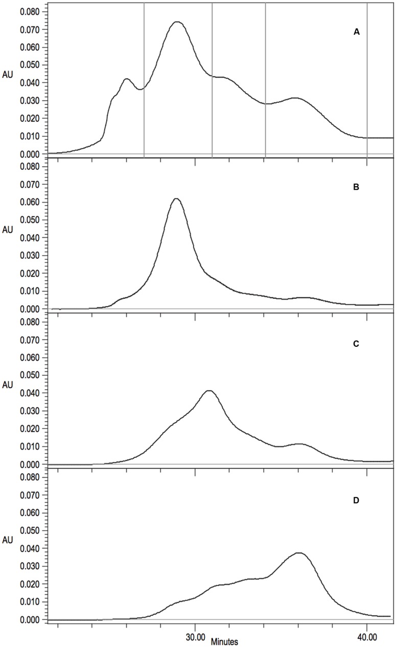
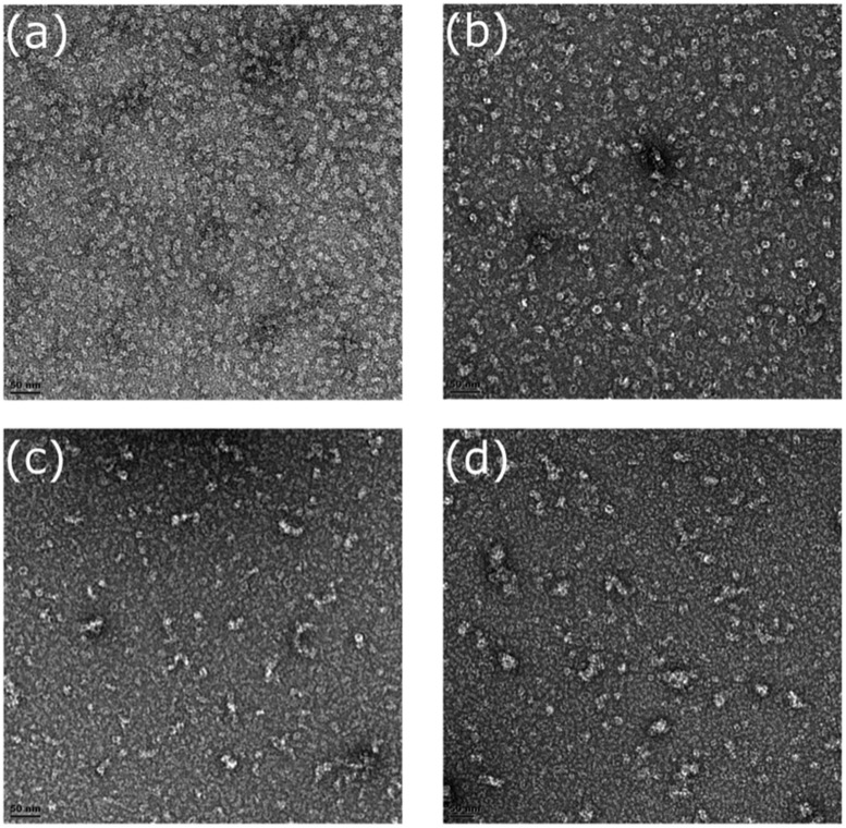
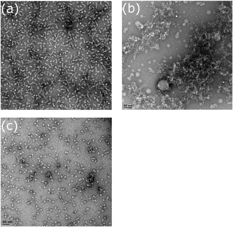
Nitrilases are of significant interest both due to their potential for industrial production of valuable products as well as degradation of hazardous nitrile-containing wastes. All known functional members of the nitrilase superfamily have an underlying dimer structure. The true nitrilases expand upon this basic dimer and form large spiral or helical homo-oligomers. The formation of this larger structure is linked to both the activity and substrate specificity of these nitrilases. The sequences of the spiral nitrilases differ from the non-spiral forming homologs by the presence of two insertion regions. Homology modeling suggests that these regions are responsible for associating the nitrilase dimers into the oligomer. Here we used cysteine scanning across these two regions, in the spiral forming nitrilase cyanide dihydratase from Bacillus pumilus (CynD), to identify residues altering the oligomeric state or activity of the nitrilase. Several mutations were found to cause changes to the size of the oligomer as well as reduction in activity. Additionally one mutation, R67C, caused a partial defect in oligomerization with the accumulation of smaller oligomer variants. These results support the hypothesis that these insertion regions contribute to the unique quaternary structure of the spiral microbial nitrilases.

Keywords: bioremediation; cyanide; cyanide dihydratase; nitrilase; oligomerization surface; quaternary structure.


Figures

FIGURE 1
FIGURE 1
Diagram showing the locations of the associating surfaces within spirals formed by members to the nitrilase superfamily. Monomers are represented by rectangles. These monomers associate to form dimers at the A-surface in all superfamily members. The dimers associate to form helical assemblies by associating across the C-surface. Two dimers are shown per turn in (A). In some superfamily members such as CynD interactions occur across the groove leading to the formation of the D-surface between adjacent turns of the spiral. In CynD, alternative interactions across the groove leading to the formation of the E-surface (B) cause to the termination of the spirals after a fixed number of dimers.
FIGURE 2
FIGURE 2
Multiple sequence alignment of: cyanide dihydratase from Bacillus pumilus C1 and Pseudomonas stutzeri (CynDpum, CynDstut) (Meyers et al., 1993; Jandhyala et al., 2003; Sewell et al., 2003), oxy-nitrilase from Synechocystis PCC6803 (3WUY) (Zhang et al., 2014), nitrilase from R. rhodochrous J1 (RRJ1) (Thuku et al., 2007), C-shaped β-alanine-synthase (βaS) from Drosophila melanogaster (2VHI) (Lundgren et al., 2008), and non spiral-forming crystallized nitrilase superfamily member 2PLQ (Nakai et al., 2000; Pace et al., 2000; Wang et al., 2001; Kumaran et al., 2003; Kimani et al., 2007). Sequence insertion regions in the spiral-forming nitrilases proposed to participate in the C-surface interaction leading to spiral formation are highlighted. Region 1 is highlighted in red and region 2 is highlighted in green. The putative catalytic residues are outlined. The sequence highlighted in blue also forms part of the interface and contributes a glutamate to the active site. The multiple sequence alignment was constructed with ClustalW2 (Larkin et al., 2007; Goujon et al., 2010). Alignment was edited and exported using Jalview software (Waterhouse et al., 2009).
FIGURE 3
FIGURE 3
The relationship between the region 1 and region 2 insertions and the C-surface based on crystal structures of two spiral forming members of the nitrilase superfamily (A) β-alanine synthase from Drosophila melanogaster (PDB id: 2VHI) and (B) the oxy-nitrilase from Synechocystis sp. PCC6803 (PDB id: 3WUY). Region 1 is highlighted in red and region 2 is highlighed in green. In both cases there is a further contributor to the interface depicted in blue. This sequence contains a glutamate (depicted in pink) that is hydrogen bonded to the lysine of the putative catalytic triad comprising a cysteine (yellow), a glutamate (pink), and the lysine (purple).
FIGURE 4
FIGURE 4
Stereoview of the model of CynD based on the structure of the oxy-nitrilase from Synechocystis sp. PCC6803 (PDB id: 3WUY). The ribbon depicting region 1 is colored red. This region includes three residues that when mutated to cysteine, caused CynD to lose activity: E64, R67, and Y70 (colored purple). It also included seven residues that when mutated to cysteine caused the activity to drop below 50% of the “wt” activity: P55, F57, Y65, T66, F69, H71, and E72 (colored pink). The ribbon depicting region 2 is colored green. It includes two residues that when mutated to cysteine caused the activity to drop below 50% of the “wt” activity: N230 and E235 (colored pink). Residue Q228, mutation of which caused aggregation of the short spirals without loss of activity is colored brown.
FIGURE 5
FIGURE 5
Activity of purified CynD proteins relative to “wt” CynD and buffer only controls (C) measured by the picric acid CN assay. Activity for each substitution mutant in the C-surface regions 1 (A, top) and 2 (B, bottom). Error bars show standard deviation from three samples.
FIGURE 6
FIGURE 6
Gel filtration analysis on Superdex 200 10/300 GL of purified CynD protein in 100 mM MOPS pH 7.7. Elution monitored as absorbance at 220 nm. Vertical gray lines highlight prominent peaks. Continuous black line indicates wild-type CynD elution peak. Representative elution patterns of substitution mutants in CynD; (A) wild type 18-mer CynD, (B) intermediate 16-mer mutants (F69C shown) (see Table 2), (C) P. stutzeri CynD 14-mer-like F57C, (D) multiple elution peaks of R67C (also see Figure 6A), (E) sloping void peak of Q228C.
FIGURE 7
FIGURE 7
(A) Gel filtration analysis and fraction collection on Superdex 200 10/300 GL of CynD R67C protein. Collection periods shown by vertical lines. (B-D) Gel filtration of concentrated fractions; (B) 14-mer like 27-31 min, (C) intermediate decamer-hexamer range 31-34 min, (D) dimer 34-40 min.
FIGURE 8
FIGURE 8
Fractions of CynD R67C from gel filtration incubated at pH 8.0, stained with 2% uranyl acetate and examined by transmission electron microscopy. (A,B) Represent early fraction and (C,D) represent later fractions.
FIGURE 9
FIGURE 9
Fractions of (A) CynD Y70C, (B) Q228C, and (C) wild type from gel filtration incubated at pH 8.0, stained with 2% uranyl acetate and examined by transmission electron microscopy.

References

    1. Abou Nader M. (2012). Directed Evolution of Cyanide Degrading Enzymes. Ph.D. dissertation, Texas A&M University, College Station, TX.
    1. Akcil A. (2003). Destruction of cyanide in gold mill effluents: biological versus chemical treatments. Biotechnol. Adv. 21 501–511. 10.1016/S0734-9750(03)00099-5 - DOI - PubMed
    1. Crum M. A., Park J. M., Sewell B. T., Benedik M. J. (2015). C-terminal hybrid mutant of Bacillus pumilus cyanide dihydratase dramatically enhances thermal stability and pH tolerance by reinforcing oligomerization. J. Appl. Microbiol. 118 881–889. 10.1111/jam.12754 - DOI - PubMed
    1. Dent K. C., Weber B. W., Benedik M. J., Sewell B. T. (2009). The cyanide hydratase from Neurospora crassa forms a helix which has a dimeric repeat. Appl. Microbiol. Biotechnol. 82 271–278. 10.1007/s00253-008-1735-4 - DOI - PubMed
    1. Gong J. S., Lu Z. M., Li H., Shi J. S., Zhou Z. M., Xu Z. H. (2012). Nitrilases in nitrile biocatalysis: recent progress and forthcoming research. Microb. Cell Fact. 11 142 10.1186/1475-2859-11-142 - DOI - PMC - PubMed

LinkOut - more resources