Incidence of Platelet Dysfunction by Thromboelastography-Platelet Mapping in Children Supported with ECMO: A Pilot Retrospective Study
- PMID: 26779465
- PMCID: PMC4702183
- DOI: 10.3389/fped.2015.00116
Incidence of Platelet Dysfunction by Thromboelastography-Platelet Mapping in Children Supported with ECMO: A Pilot Retrospective Study
Abstract
Background: Bleeding complications are common and decrease the odds of survival in children supported with extracorporeal membrane oxygenation (ECMO). The role of platelet dysfunction on ECMO-induced coagulopathy and resultant bleeding complications is not well understood. The primary objective of this pilot study was to determine the incidence and magnitude of platelet dysfunction according to thromboelastography (TEG(®))-platelet mapping (PM) testing.
Methods: Retrospective chart review of children <18 years old who required ECMO at a tertiary level hospital. We collected TEG(®)-PM and conventional coagulation tests data. We also collected demographic, medications, blood products administered, and clinical outcome data. We defined severe platelet dysfunction as <50% aggregation in response to an agonist.
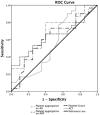
Results: We identified 24 out of 46 children on ECMO, who had TEG(®)-PM performed during the study period. We found the incidence of severe bleeding was 42% and mortality was 54% in our study cohort. In all samples measured, severe qualitative platelet dysfunction was more common for adenosine diphosphate (ADP)-mediated aggregation (92%) compared to arachidonic acid (AA)-mediated aggregation (75%) (p = 0.001). Also, ADP-mediated percent of platelet aggregation was significant lower than AA-mediated platelet aggregation [15% (interquartile range, IQR 2.8-48) vs. 49% (IQR 22-82.5), p < 0.001]. There was no difference in kaolin-activated heparinase TEG(®) parameters between the bleeding group and the non-bleeding group. Only absolute platelet count and TEG(®)-PM had increased predictive value on receiver operating characteristics analyses for severe bleeding and mortality compared to activated clotting time.
Conclusion: We found frequent and severe qualitative platelet dysfunction on TEG(®)-PM testing in children on ECMO. Larger studies are needed to determine if the assessment of qualitative platelet function by TEG(®)-PM can improve prediction of bleeding complications for children on ECMO.
Keywords: ECMO; ECMO-induced coagulopathy; anticoagulation; heparin; platelet dysfunction; platelet mapping; thromboelastography.
Figures


References
Grants and funding
LinkOut - more resources
Full Text Sources
Other Literature Sources

