

Review
doi: 10.5339/gcsp.2015.44.
eCollection 2015.
Current management of coarctation of the aorta
Affiliations
- PMID: 26779519
- PMCID: PMC4710863
- DOI: 10.5339/gcsp.2015.44
Item in Clipboard
Review
Current management of coarctation of the aorta
Glob Cardiol Sci Pract.
.
Abstract
Coarctation of the aorta (C) is the sixth most common lesion in congenital heart disease and represents a spectrum of aortic narrowing that varies from a discrete entity to tubular hypoplasia. This condition was once thought to be a relatively simple lesion that would be "cured" upon repair of the narrowing, however, despite relief of the anatomical obstruction the subsequent risk of early morbidity and death persists. This review outlines the optimal management strategy of this disease from neonatal to adult life and provides insights to approach this straightforward but challenging condition.
Figures

Survival curves to 30 years of 588 surgically treated patients (solid line) and the expected survival of an age and sex-matched population based on cohort life tables (dashed line).
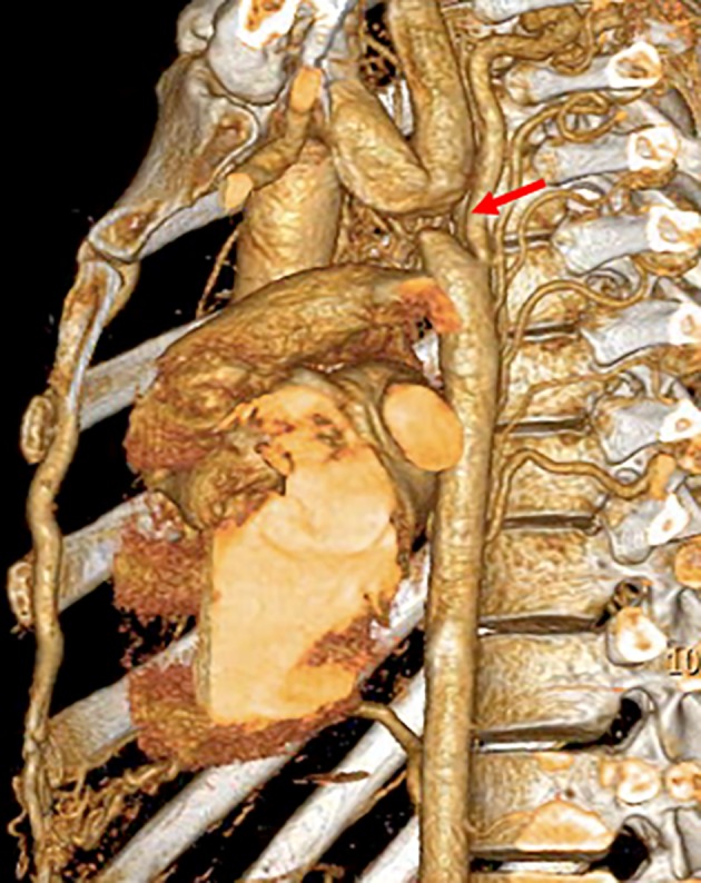
Computed Tomography (CT) reconstruction demonstrating aortic coarctation (arrow).
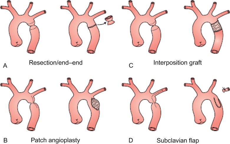
Major surgical aortic coarctation repair techniques.
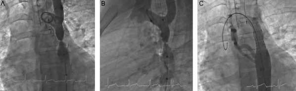
Stenting of tight native coarctation. Native coarctation with multiple collaterals in A) LAO and B) straight lateral views. C) No residual stenosis following stent implantation.

A) Aortic angiogram demonstrating large aneurysm at the site of previously repaired coarctation B) Angiogram following successful aneurysm repair using endovascular stent graft.

Angiogram of a young patient with coarctation and cerebral aneurysm (arrow).
Similar articles
-
Coarctation of the aorta: evaluation and management.Curr Opin Cardiol. 2009 Nov;24(6):509-15. doi: 10.1097/HCO.0b013e328330cc22. Curr Opin Cardiol. 2009. PMID: 19667980 Review.
-
Coarctation of the aorta: from fetal life to adulthood.Cardiol J. 2011;18(5):487-95. doi: 10.5603/cj.2011.0003. Cardiol J. 2011. PMID: 21947983 Review.
-
Echocardiography of coarctation of the aorta, aortic arch hypoplasia, and arch interruption: strategies for evaluation of the aortic arch.Cardiol Young. 2016 Dec;26(8):1553-1562. doi: 10.1017/S1047951116001670. Cardiol Young. 2016. PMID: 28148317 Review.
-
[; SURGICAL TREATMENT OF TRANSPOSITION OF THE GREAT ARTERIES AND AORTIC ARCH HYPOPLASIA].Georgian Med News. 2020 Jul-Aug;(304-305):85-90. Georgian Med News. 2020. PMID: 32965255 Russian.
-
[Long-term results after surgery of coarctation of the aorta in neonates and children].Arch Mal Coeur Vaiss. 1997 Dec;90(12 Suppl):1723-8. Arch Mal Coeur Vaiss. 1997. PMID: 9587457 Review. French.
Cited by
-
Aortic Arch Variants and Anomalies: Embryology, Imaging Findings, and Clinical Considerations.J Cardiovasc Imaging. 2022 Oct;30(4):231-262. doi: 10.4250/jcvi.2022.0058. J Cardiovasc Imaging. 2022. PMID: 36280266 Free PMC article. Review.
-
Aortic coarctation in an asymptomatic patient with arterial hypertension initially evaluated by Doppler ultrasound in the emergency room: A case report.Radiol Case Rep. 2022 May 30;17(8):2689-2692. doi: 10.1016/j.radcr.2022.04.043. eCollection 2022 Aug. Radiol Case Rep. 2022. PMID: 35663808 Free PMC article.
-
Coarctation of the Aorta with Aortic Arch Hypoplasia: Midterm Outcomes of Aortic Arch Reconstruction with Autologous Pulmonary Artery Patch.Chin Med J (Engl). 2017 Dec 5;130(23):2802-2807. doi: 10.4103/0366-6999.215279. Chin Med J (Engl). 2017. PMID: 28936993 Free PMC article.
-
Necessity of life-long follow-up after surgery for coarctation of the aorta: a case series of very late false aneurysm formation.Eur Heart J Case Rep. 2022 Feb 16;6(2):ytac073. doi: 10.1093/ehjcr/ytac073. eCollection 2022 Feb. Eur Heart J Case Rep. 2022. PMID: 35233500 Free PMC article.
-
Stanford type B aortic dissection in an elderly patient with silent aortic coarctation.Tzu Chi Med J. 2017 Jul-Sep;29(3):183-184. doi: 10.4103/tcmj.tcmj_65_17. Tzu Chi Med J. 2017. PMID: 28974916 Free PMC article. No abstract available.
References
-
- Hoffman JI, Kaplan S. The incidence of congenital heart disease. Journal of the American College of Cardiology. 2002;39(12):1890–1900. Epub 2002/06/27. - PubMed
-
- Warnes CA, Williams RG, Bashore TM, Child JS, Connolly HM, Dearani JA. ACC/AHA 2008 Guidelines for the Management of Adults with Congenital Heart Disease: a report of the American College of Cardiology/American Heart Association Task Force on Practice Guidelines (writing committee to develop guidelines on the management of adults with congenital heart disease) Circulation. 2008;118(23):e714–e833. et al. Epub 2008/11/11. - PubMed
-
- Jenkins NP, Ward C. Coarctation of the aorta: natural history and outcome after surgical treatment. QJM: monthly journal of the Association of Physicians. 1999;92(7):365–371. Epub 2000/01/11. - PubMed
-
- Kaushal S, Backer CL, Patel JN, Patel SK, Walker BL, Weigel TJ. Coarctation of the aorta: midterm outcomes of resection with extended end-to-end anastomosis. The Annals of thoracic surgery. 2009;88(6):1932–1938. et al. Epub 2009/11/26. - PubMed
Publication types
LinkOut - more resources
Full Text Sources
Other Literature Sources